

In vitro studies on the effect of particle size on macrophage responses to nanodiamond wear debris
- PMID: 22342422
- PMCID: PMC3314099
- DOI: 10.1016/j.actbio.2012.01.033
In vitro studies on the effect of particle size on macrophage responses to nanodiamond wear debris
Abstract
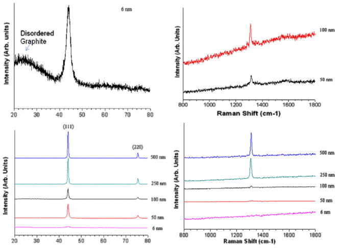
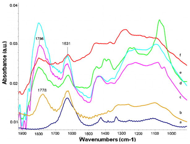
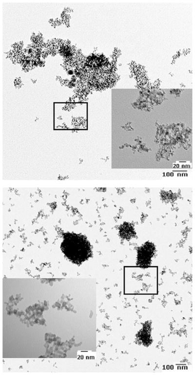
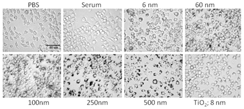
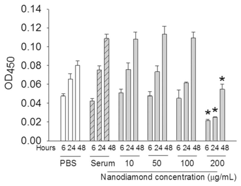
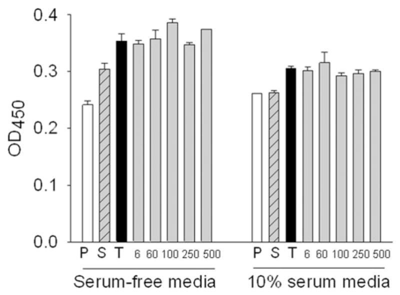
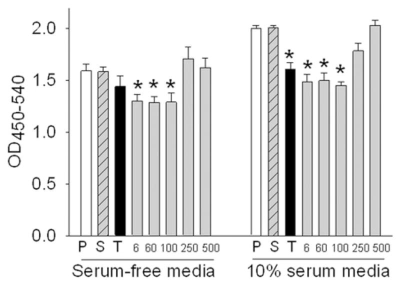
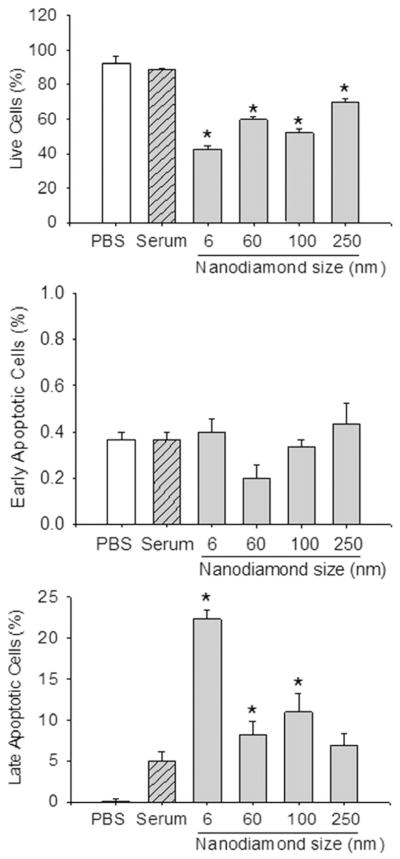
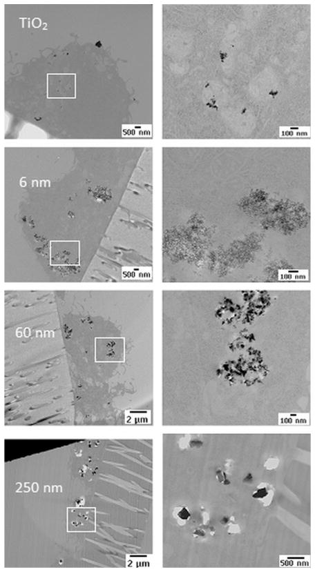
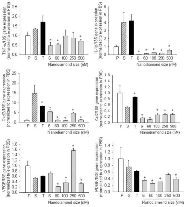
Nanostructured diamond coatings improve the smoothness and wear characteristics of the metallic component of total hip replacements and increase the longevity of these implants, but the effect of nanodiamond wear debris on macrophages needs to be determined to estimate the long-term inflammatory effects of wear debris. The objective was to investigate the effect of the size of synthetic nanodiamond particles on macrophage proliferation (BrdU incorporation), apoptosis (Annexin-V flow cytometry), metabolic activity (WST-1 assay) and inflammatory cytokine production (qPCR). RAW 264.7 macrophages were exposed to varying sizes (6, 60, 100, 250 and 500 nm) and concentrations (0, 10, 50, 100 and 200 μg ml(-1)) of synthetic nanodiamonds. We observed that cell proliferation but not metabolic activity was decreased with nanoparticle sizes of 6-100 nm at lower concentrations (50 μg ml(-1)), and both cell proliferation and metabolic activity were significantly reduced with nanodiamond concentrations of 200 μg ml(-1). Flow cytometry indicated a significant reduction in cell viability due to necrosis irrespective of particle size. Nanodiamond exposure significantly reduced gene expression of tumor necrosis factor-α, interleukin-1β, chemokine Ccl2 and platelet-derived growth factor compared to serum-only controls or titanium oxide (anatase 8 nm) nanoparticles, with variable effects on chemokine Cxcl2 and vascular endothelial growth factor. In general, our study demonstrates a size and concentration dependence of macrophage responses in vitro to nanodiamond particles as possible wear debris from diamond-coated orthopedic joint implants.
Copyright © 2012 Acta Materialia Inc. Published by Elsevier Ltd. All rights reserved.
Figures
References
-
- American Academy of Orthopaedic Surgeons. 2006 < http://www.aaos.org/>.
-
- Amstutz HC, Campbell P, Kossovsky N, Clarke IC. Mechanism and clinical significance of wear debris induced osteolysis. Clin Orthop Relat Res. 1992;276:7–18. - PubMed
-
- Goldring SR, Schiller AL, Roelke M, Rourke CM, O’Neil DA, Harris WH. The synovial-like membrane at the bone-cement interface in loose total hip replacements and its proposed role in bone lysis. J Bone Joint Surg Am. 1983;65(5):575–84. - PubMed
-
- Mercuri LG. The TMJ concepts patient fitted total temporomandibular joint reconstruction prosthesis in total temporomandibular joint reconstruction. Oral Maxillofac Surg Clin North Am. 2000;12:73–91.
-
- Amstutz HC, Grigoris P. Metal on metal bearings in hip arthroplasty. Clin Orthop Relat Res. 1996;329:S11–34. - PubMed
Publication types
MeSH terms
Substances
Grants and funding
LinkOut - more resources
Full Text Sources
