. 2012 Feb 24;36(2):239-50.
doi: 10.1016/j.immuni.2011.12.015.

Suppression of cytokine signaling by SOCS3: characterization of the mode of inhibition and the basis of its specificity

Affiliations

Suppression of cytokine signaling by SOCS3: characterization of the mode of inhibition and the basis of its specificity

Jeffrey J Babon et al. Immunity. .

Abstract

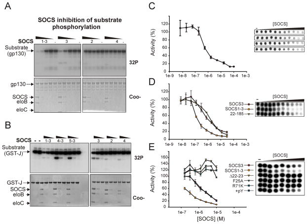
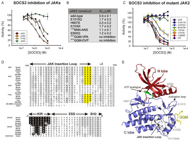
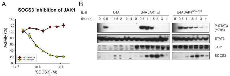
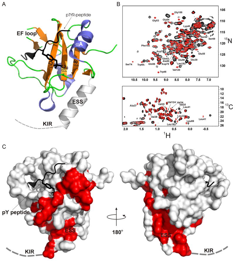
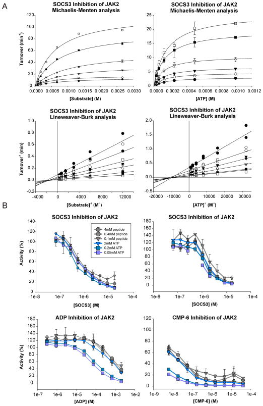
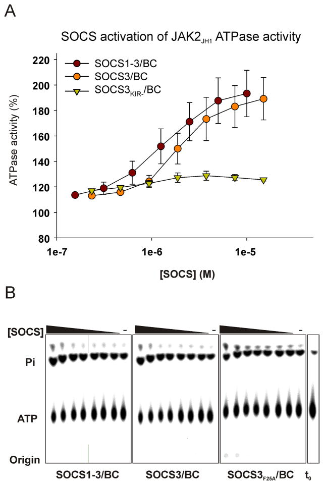
Janus kinases (JAKs) are key effectors in controlling immune responses and maintaining hematopoiesis. SOCS3 (suppressor of cytokine signaling-3) is a major regulator of JAK signaling and here we investigate the molecular basis of its mechanism of action. We found that SOCS3 bound and directly inhibited the catalytic domains of JAK1, JAK2, and TYK2 but not JAK3 via an evolutionarily conserved motif unique to JAKs. Mutation of this motif led to the formation of an active kinase that could not be inhibited by SOCS3. Surprisingly, we found that SOCS3 simultaneously bound JAK and the cytokine receptor to which it is attached, revealing how specificity is generated in SOCS action and explaining why SOCS3 inhibits only a subset of cytokines. Importantly, SOCS3 inhibited JAKs via a noncompetitive mechanism, making it a template for the development of specific and effective inhibitors to treat JAK-based immune and proliferative diseases.


Figures

Figure 1
Figure 1. SOCS3 and SOCS1-3 inhibit substrate phosphorylation by JAK2
(A) Autoradiography (upper panel) and Coomassie stained (lower panel) SDS-PAGE analysis of JAK2JH1 catalyzed phosphorylation of the gp130 cytoplasmic domain in the presence of serial 10-fold dilutions of SOCS-elonginBC complexes. Both SOCS3 and a SOCS1-3 chimaera were effective inhibitors of substrate phosphorylation. Gradient bars represent decreasing concentration of each SOCS construct. (10μM, 1μM, 100nM, 10nM, left to right) (B) As in (A) except a different substrate (GST-J) was used as a substrate. SOCS concentration was 5μM, 1μM, 200nM, left to right in each case. (C) Quantitative kinase inhibition experiments. As in (A) except reactions were spotted onto nitrocellulose filters. The experiment was performed in quadruplicate, raw data shown above and quantitated below. The control reaction (0 SOCS) was normalized to 100%. (D) SOCS1-3 is a more effective inhibitor of JAK2 than SOCS3. As in (C) except a STAT5b peptide was used as substrate and the reaction spotted onto P81 phosphocellulose paper. Reactions contained 30, 12, 5, 2, 0.8, 0.3, 0.1, 0.04, 0.02, 0.008, 0 μM SOCS (left to right). (E) SOCS3 constructs that contained mutations in the KIR (F25A), SH2 domain (R71K) or that lacked the first eight residues of the KIR (Δ22–29) did not inhibit JAK2. As in (D). Reactions contained 10, 5, 2.5, 1.2, 0.6, 0.3, 0.15, 0 μM SOCS (left to right).. The addition of gp130 phosphopeptide (pY) did not affect inhibition. Error bars represent +/− range of two experiments. Data are normalized to no-inhibitor controls
Figure 2
Figure 2. SOCS3 inhibits JAK1, JAK2 and TYK2 but not JAK3 due to a three residue motif in the JAK insertion loop
(A) Kinase inhibition assays were performed using the kinase domain of all four JAKs. Only JAK3JH1 was not inhibited by SOCS3. Error bars represent +/− range from two experiments. Data were normalized to no-inhibitor controls (B) Mutating the GQM motif makes JAK2 non-responsive to SOCS3. All other mutations have no effect. (C) G1071 and M1073 are absolutely required for SOCS3 inhibition. As in (A) except point mutants of JAK2 were tested. (D) Sequence alignment of the GQM region of all JAKs and the KIR of SOCS3 and SOCS1. Highly conserved residues are shown boxed in grey, the GQM motif in yellow, conserved residues in the KIR in black. (E) The structure of JAK2, PDB ID 2B7A. The GQM motif is solvent exposed and shown in yellow. The location of mutated residues from (B) are indicated by grey arrows. *Zebrafish JAK2b is grouped with TYK2 in this figure.
Figure 3
Figure 3. IL-6 induced phosphorylation of STAT3 is prolonged in the presence of JAK1GQM-DVP
(A) Kinase inhibition assays were performed using the kinase domain of JAK1 and JAK1GQM-DVP. JAK1GQM-DVP was not inhibited by SOCS3. Error bars represent +/− range from two experiments. Data were normalized to no-inhibitor controls. (B) Wild-type and mutant (GQM-DVP) JAK1 constructs were transfected into JAK1−/− (U4A) cells. Cells were stimulated with IL-6 plus s-IL-6Rα for 15 min and then harvested at the indicated times. Lysates were analyzed by immunoblotting with antibodies specific for phosphorylated STAT3, total STAT3, SOCS3 and JAK1.
Figure 4
Figure 4. NMR analysis identifies the surface of SOCS3 that interacts with JAK2
(A) Ribbon diagram of the structure of SOCS322–185 (PDB ID 2HMH) with important secondary structural motifs indicated. The dashed line indicates the first seven residues of the KIR which are unstructured in the absence of JAK. (B) 1H-15N and 1H-13C HMQC analysis of the SOCS3-JAK2JH1 interaction are shown in upper and lower panels respectively. The SOCS spectra are in red and the SOCS3-JAK2JH1 spectrum in black. (C) Surface diagram of SOCS3. Residues whose resonances shift in the presence of JAK2JH1 highlighted in red. The orientation of SOCS3 on the left is identical to that in (A).
Figure 5
Figure 5. SOCS3 is a non-competitive inhibitor of JAK2
(A) Michaelis-Menten (upper panels) and Lineweaver-Burk (lower panels) analysis of SOCS3 inhibition of JAK2JH1. ATP (left) and peptide substrate (right) titrations show that SOCS3 alters Vmax but not Km, indicating non-competitive inhibition. Inhibition experiments were performed using 10nM JAK and 0–8μM SOCS3 with varying concentrations of ATP and substrate. Lineweaver-Burk curves that intersect on the abscissa indicate non-competitive inhibition. Error bars represent +/− range from two experiments. (B) JAK2 inhibition experiments were performed in the presence of SOCS3 (upper panels, 2 independent experiments) or in the presence of ATP competitive inhibitors ADP and CMP-6 (lower panels). The IC50 values of SOCS3 were unaffected by ATP concentration unlike the IC50 values of ADP and CMP-6. Error bars represent +/− range from two experiments. Data are normalized to no-inhibitor controls.
Figure 6
Figure 6. SOCS3 has an activating effect on JAK2 JH1 ATPase activity
Assays were analyzed by TLC (lower panels) and quantified (upper panel). In control reactions (right hand lane of each panel) JAK2JH1 hydrolysed ATP at 0.05 s−1 under these conditions compared to 0.09 s−1 in the presence of 15μM SOCS3. Error bars: +/− range, two experiments. Data are normalized to no inhibitor controls.

Comment in

References

    1. Adams JA. Kinetic and catalytic mechanisms of protein kinases. Chem Rev. 2001;101:2271–2290. - PubMed
    1. Babon JJ, McManus EJ, Yao S, DeSouza DP, Mielke LA, Sprigg NS, Willson TA, Hilton DJ, Nicola NA, Baca M, et al. The structure of SOCS3 reveals the basis of the extended SH2 domain function and identifies an unstructured insertion that regulates stability. Mol Cell. 2006;22:205–216. - PubMed
    1. Babon JJ, Sabo JK, Soetopo A, Yao S, Bailey MF, Zhang JG, Nicola NA, Norton RS. The SOCS box domain of SOCS3: structure and interaction with the elonginBC-cullin5 ubiquitin ligase. J Mol Biol. 2008;381:928–940. - PMC - PubMed
    1. Babon JJ, Sabo JK, Zhang JG, Nicola NA, Norton RS. The SOCS box encodes a hierarchy of affinities for Cullin5: implications for ubiquitin ligase formation and cytokine signalling suppression. J Mol Biol. 2009;387:162–174. - PMC - PubMed
    1. Babon JJ, Yao S, DeSouza DP, Harrison CF, Fabri LJ, Liepinsh E, Scrofani SD, Baca M, Norton RS. Secondary structure assignment of mouse SOCS3 by NMR defines the domain boundaries and identifies an unstructured insertion in the SH2 domain. Febs J. 2005;272:6120–6130. - PubMed

Publication types

MeSH terms