

Development of functional imaging in the human brain (fMRI); the University of Minnesota experience
- PMID: 22342875
- PMCID: PMC3530260
- DOI: 10.1016/j.neuroimage.2012.01.135
Development of functional imaging in the human brain (fMRI); the University of Minnesota experience
Abstract
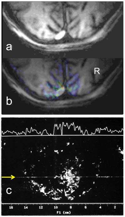
The human functional magnetic resonance imaging (fMRI) experiments performed in the Center for Magnetic Resonance Research (CMRR), University of Minnesota, were planned between two colleagues who had worked together previously in Bell Laboratories in the late nineteen seventies, namely myself and Seiji Ogawa. These experiments were motivated by the Blood Oxygenation Level Dependent (BOLD) contrast developed by Seiji. We discussed and planned human studies to explore imaging human brain activity using the BOLD mechanism on the 4 Tesla human system that I was expecting to receive for CMRR. We started these experiments as soon as this 4 Tesla instrument became marginally operational. These were the very first studies performed on the 4 Tesla scanner in CMRR; had the scanner become functional earlier, they would have been started earlier as well. We were aware of the competing effort at the Massachusetts General Hospital (MGH) and we knew that they had been informed of our initiative in Minneapolis to develop fMRI. We had positive results certainly by August 1991 annual meeting of the Society of Magnetic Resonance in Medicine (SMRM). I believe, however, that neither the MGH colleagues nor us, at the time, had enough data and/or conviction to publish these extraordinary observations; it took more or less another six months or so before the papers from these two groups were submitted for publication within five days of each other to the Proceedings of the National Academy of Sciences, USA, after rejection by Nature in our case. Thus, fMRI was achieved independently and at about the same time at MGH, in an effort credited largely to Ken Kwong, and in CMRR, University of Minnesota in an effort led by myself and Seiji Ogawa.
Copyright © 2012 Elsevier Inc. All rights reserved.
Figures
References
-
- Biswal B, Yetkin FZ, Haughton VM, Hyde JS. Functional connectivity in the motor cortex of resting human brain using echo-planar MRI. Magnetic Resonance in Medicine. 1995;34(4):537–541. - PubMed
-
- Cheng K, Waggoner RA, Tanaka K. Human ocular dominance columns as revealed by high-field functional magnetic resonance imaging. Neuron. 2001;32(2):359–374. - PubMed
-
- Duong TQ, Silva AC, Lee SP, Kim SG. Functional MRI of calcium-dependent synaptic activity: cross correlation with CBF and BOLD measurements [In Process Citation] Magn Reson Med. 2000;43(3):383–392. - PubMed
Publication types
MeSH terms
Substances
Grants and funding
LinkOut - more resources
Full Text Sources
Medical
