Review
. 2013 Jun:69:3-15.
doi: 10.1016/j.neuropharm.2012.01.022. Epub 2012 Feb 4.

Defining "epileptogenesis" and identifying "antiepileptogenic targets" in animal models of acquired temporal lobe epilepsy is not as simple as it might seem

Affiliations
Review

Defining "epileptogenesis" and identifying "antiepileptogenic targets" in animal models of acquired temporal lobe epilepsy is not as simple as it might seem

Robert S Sloviter et al. Neuropharmacology. 2013 Jun.

Abstract

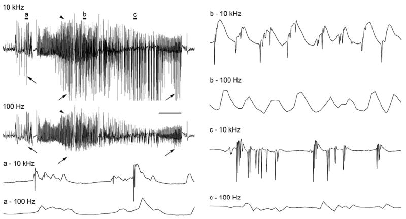
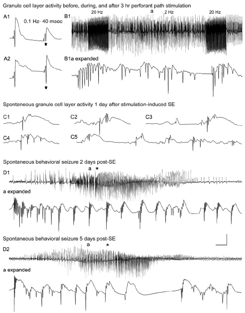
The "latent period" between brain injury and clinical epilepsy is widely regarded to be a seizure-free, pre-epileptic state during which a time-consuming cascade of molecular events and structural changes gradually mediates the process of "epileptogenesis." The concept of the "latent period" as the duration of "epileptogenesis" implies that epilepsy is not an immediate result of brain injury, and that anti-epileptogenic strategies need to target delayed secondary mechanisms that develop sometime after an initial injury. However, depth recordings made directly from the dentate granule cell layers in awake rats after convulsive status epilepticus-induced injury have now shown that whenever perforant pathway stimulation-induced status epilepticus produces extensive hilar neuron loss and entorhinal cortical injury, hyperexcitable granule cells immediately generate spontaneous epileptiform discharges and focal or generalized behavioral seizures. This indicates that hippocampal injury caused by convulsive status epilepticus is immediately epileptogenic and that hippocampal epileptogenesis requires no delayed secondary mechanism. When latent periods do exist after injury, we hypothesize that less extensive cell loss causes an extended period during which initially subclinical focal seizures gradually increase in duration to produce the first clinical seizure. Thus, the "latent period" is suggested to be a state of "epileptic maturation," rather than a prolonged period of "epileptogenesis," and therefore the antiepileptogenic therapeutic window may only remain open during the first week after injury, when some delayed cell death may still be preventable. Following the perhaps unavoidable development of the first focal seizures ("epileptogenesis"), the most fruitful therapeutic strategy may be to interrupt the process of "epileptic maturation," thereby keeping focal seizures focal. This article is part of the Special Issue entitled 'New Targets and Approaches to the Treatment of Epilepsy'.


Figures

Figure 1
Figure 1
Brain structure 3 days after status epilepticus induced by systemic injection of kainic acid (KA; 12 mg/kg sc). In some animals given kainate or pilocarpine systemically, prolonged status epilepticus caused apparent hemorrhages in a multitude of brain structures, which were not cleared by vascular perfusion. (A) and (C): two coronal views of the same brain during the sectioning process. Note apparent hemorrhagic foci in the hippocampi, thalamus, and temporal cortices. (B) and (D): foci preferentially involve the CA1 pyramidal cell layer (arrow) and the dorsolateral thalamus (arrowhead). (E) and (F): in a Fluoro Jade B-stained section from the brain shown in (A)-(D), degenerating neurons are fluorescent. Note that the area CA1 pathology consists of a vascular expansion that is continuous with a capillary in the hippocampal fissure (hf; arrow), not an extravascular hemorrhage. (G): in a different kainate-treated rat, smaller focal vascular expansions occurred within the stratum radiatum (arrows). (H) At higher magnification, degenerating CA1 pyramidal cell somata were evident only adjacent to the vascular pathology. These results suggest that, in some cases, CA1 pyramidal cell layer injury in rats subjected to prolonged status epilepticus may be ischemic in nature, rather than excitotoxic. Abbreviations: sp: stratum pyramidale; sr: stratum radiatum; slm: stratum lacunosum-moleculare; hf: hippocampal fissure. Magnifications: 5X (A and C); 13.5X (B and D); 22X (E and G); 55X (F and H). From Sloviter, 2005.
Figure 2
Figure 2
Evoked and spontaneous granule cell activity recorded from the granule cell layers of chronically epileptic, pilocarpine treated-rats. A: Granule cell layer seizure discharges evoked in a control animal (60 days post-saline treatment) by perforant path stimulation at 1 Hz (with paired-pulses 40 msec apart) for 40 sec. Note that evoked granule cell activity generated both low-frequency, positive-going field potentials (expanded box 1) and high-frequency, negative-going population spikes (expanded box 2). B: In an epileptic animal 2 days post-SE, spontaneous granule cell layer activity recorded during a spontaneous behavioral seizure exhibited both positive-going field potentials (expanded box 1) and negative-going epileptiform discharges (expanded box 2) that were qualitatively similar to those evoked in the control rat in A. C: Thirteen days later, spontaneous granule cell layer activity still exhibited high-frequency, negative-going epileptiform discharges. D: Forty-seven days after SE, the spontaneous granule cell layer activity recorded in the same animal during a spontaneous seizure exhibited single population spikes (expanded box 1) and positive-going field potentials (expanded boxes 2 and 3) but no seizure discharges (repetitive population spikes). Note that granule cells generated only single population spikes ~7 seconds after the onset of the observed behavioral seizure (expanded box 1) and then generated only field potentials for the duration of the behavioral seizure. E: Evoked granule cell seizure afterdischarges in the same chronically epileptic, pilocarpine-treated rat 45 days post-SE (tested 2 days prior to the activity shown in D), illustrating that, despite the lack of spontaneous seizure discharges in the chronic epileptic state, abnormally high-frequency (4 –10Hz) stimulation could evoke granule cell seizure discharges (expanded box 1), as well as positive-going field potentials (expanded box 2). The absence of seizure discharges recorded from the granule cell layers during spontaneous behavioral seizures was a consistent observation in epileptic animals subsequently shown to have dentate hilar cell loss and mossy fiber sprouting. Calibration bars: 3.5 mV, 30 msec in A; 5 mV, 2 sec for B; 4 mV, 30 msec for B1,2; 2.5 mV, 2 sec for C; 3 mV, 100 msec for C1–3; 3 mV, 2 sec for D; 4 mV, 25 msec for D1,2. From Harvey and Sloviter, 2005.
Figure 3
Figure 3
A–C: Quantitative frequency analysis of “early” and “late” spontaneous seizures recorded in the same chronically epileptic, pilocarpine-treated rats. A1,2: Joint time frequency analysis spectrograms of “early” (2 days post-SE) and “late” (47 days post-SE) seizures in the same awake, epileptic animal, indicating when granule cell layer activities of different frequencies occurred in relation to the behavioral seizure onsets. Note that all high-frequency components (100 – 600 Hz) were greater in magnitude (darkness and height of peaks) in the early vs. late post-status epilepticus (SE) states, indicating a loss of epileptiform discharges over time. Also note that these high-frequency components occurred after, rather than before, the spontaneous behavioral seizure onsets (onset of forepaw clonus and rearing). B1: Averaged power spectrum of six randomly selected, 1-minute-long traces of granule cell activity during SE. B2,3: The six first and last spontaneous seizures during the “early” and “late” spontaneous seizures, respectively, in a typical epileptic pilocarpine- treated rat. Note that all frequencies between 100 and 600 Hz were higher during “early” seizures (B2) than during “late” seizures (B3), indicating the loss of high-frequency epileptiform granule cell discharges in the chronic epileptic state. C1: The average integral taken from the averaged power spectrum (100 – 600 Hz) in all five chronically epileptic rats that were implanted before SE. Note the significant loss (P< 0.012) of all high-frequency components. C2,C3: The average integrals taken from the averaged power spectra of the two high-frequency bands (165–175 Hz and 340–360 Hz) in all five epileptic rats. Note the significant loss (P < 0.012 and 0.014, respectively) of both narrow frequency bands. Calibration bars: 5 mV, 4 sec. From Harvey and Sloviter, 2005.
Figure 4
Figure 4
Atypical recruitment of granule cell and CA1 pyramidal cell discharges during a spontaneous seizure in a chronically epileptic rat. A,B: In this animal, perfusion fixed 56 days post-SE, seizure discharges were recorded from both dorsal hippocampal granule cell (GCL) and CA1 pyramidal cell layers beginning approximately 25 seconds after the behavioral seizure onset. C1: c-Fos immunostaining of the dorsal hippocampus in a coronal section revealed c-Fos expression in all hippocampal neurons. D1: c-Fos immunostaining of the ventral hippocampus in a horizontal section from the same rat revealed c-Fos expression in pyramidal layer neurons but not in the dentate gyrus (sg; stratum granulosum). C2,D2: Note that Timm staining revealed less extensive mossy fiber sprouting in the dorsal hippocampus (C2) than in the ventral hippocampus (D2), and a lack of c-Fos expression in the more heavily mossy fiber-sprouted ventral dentate gyrus. Calibration bars: 10 mV, 5 seconds (compressed areas); 6 mV, 75 msec (expanded boxes). Scale bar: 400 um in D (applies to C,D). From Harvey and Sloviter, 2005.
Figure 5
Figure 5
A spontaneous electrographic seizure recorded from the dentate granule cell layer of an awake rat 2 days after 3 hr of stimulation-induced status epilepticus. This first spontaneous granule cell-onset seizure was recorded at two acquisition rates (10 kHz and 100 Hz) simultaneously to illustrate the loss of high frequency data at the lower acquisition rate. Note that presentation of the recording at a compressed timescale (top left) makes it appear that both recordings are similar superficially. However, even at this compression, it can be seen that the slower 100 Hz recording lacks the negative-going population spikes (arrows). Conversely, the slower positive-going potentials are recorded by both methods (arrowheads). Expansion of the recordings clearly shows the selective loss of all high frequency data at the 100 Hz rate, making it impossible to know if population spikes (synchronous granule cell discharges) are present. Scale bar: 4 sec in the compressed traces and 60 msec in traces a-c.
Figure 6
Figure 6
Dentate granule cell excitability and spontaneous activity before and 1–5 days after 3 hours of perforant pathway stimulation-induced convulsive status epilepticus (SE). A1: Before SE, paired-pulse perforant pathway stimulation at 0.1 Hz and an interstimulus interval of 40 msec evokes granule cell responses that exhibit partial suppression of the amplitude of the second population spike (arrow). A2: Three days after 3 hours of SE, the identical afferent stimulation failed to suppress the second population spike (arrow). B1: Granule cell layer activity during 3 hours of perforant pathway stimulation in the same awake rat. The stimulation paradigm involved continuous stimulation at 2 Hz with paired pulses delivered at a 40-msec inter- pulse interval plus 10-second-long 20-Hz trains delivered once per minute. Note the morphology of the granule cell epileptiform discharges during the 2 Hz intertrain interval (a) in the expanded trace (B1a expanded). C: On the first day after 3 hours of stimulation- induced SE, a granule cell layer electrode recorded spontaneous granule cell field “EPSPs” and population spikes that closely resemble the evoked responses in A. D: Granule cell layer activity during spontaneous behavioral seizures during the first week post-SE. D1: On day 2 post-SE, granule cell layer activity amplitude increased before the behavioral onset of the second behavioral seizure on that day (marked by asterisk). D1a (expanded): Expanded trace of the region above marked “a,” showing that the high-amplitude activity in D1 consisted of granule cell epileptiform discharges that preceded the behavioral seizure onset (asterisk). D2: Three days later, the fourth spontaneous behavioral seizure exhibited nearly identical features, including high-frequency granule cell epileptiform discharges (D2a expanded) that preceded the behavioral seizure-onset (asterisk). Calibration bars: 14 msec and 9mV in A; 7 seconds and 9 mV in B1; 46 msec and 9mV in B1 (expanded); 40 msec and 9 mV in C; 3.4 seconds and 9 mV in D1,D2; 53 msec and 9 mV in D1 (expanded); 60 msec and 9 mV in D2 (expanded). From Bumanglag and Sloviter, 2008.

References

    1. Andersen P, Holmqvist B, Voorhoeve PE. Entorhinal activation of dentate granule cells. Acta Physiol Scand. 1966;66:448–460. - PubMed
    1. Ben-Ari Y. Limbic seizure and brain damage produced by kainic acid: mechanisms and relevance to human temporal lobe epilepsy. Neuroscience. 1985;14:375–403. - PubMed
    1. Biagini G, Baldelli E, Longo D, Contri MB, Guerrini U, Sironi L, Gelosa P, Zini I, Ragsdale DS, Avoli M. Proepileptic influence of a focal vascular lesion affecting entorhinal cortex-CA3 connections after status epilepticus. J Neuropathol Exp Neurol. 2008;67:687–701. - PMC - PubMed
    1. Blümcke I, Pauli E, Clusmann H, Schramm J, Becker A, Elger C, Merschhemke M, Meencke HJ, Lehmann T, von Deimling A, Scheiwe C, Zentner J, Volk B, Romstöck J, Stefan H, Hildebrandt M. A new clinico-pathological classification system for mesial temporal sclerosis. Acta Neuropathol. 2007;113:235–244. - PMC - PubMed
    1. Bower MR, Buckmaster PS. Changes in granule cell firing rates precede locally recorded spontaneous seizures by minutes in an animal model of temporal lobe epilepsy. J Neurophysiol. 2008;99:2431–2442. - PubMed

Publication types

Substances