. 2012 May 15;355(1):121-34.
doi: 10.1016/j.mce.2012.02.001. Epub 2012 Feb 11.

Separate regions of glucocorticoid receptor, coactivator TIF2, and comodulator STAMP modify different parameters of glucocorticoid-mediated gene induction

Affiliations

Separate regions of glucocorticoid receptor, coactivator TIF2, and comodulator STAMP modify different parameters of glucocorticoid-mediated gene induction

Smita Awasthi et al. Mol Cell Endocrinol. .

Abstract

Increased specificity in steroid-regulated gene expression is a long-sought goal of endocrinologists. Considerable progress has resulted from the discovery of coactivators, corepressors, and comodulators that adjust the total activity (A(max)) of gene induction. Two less frequently quantitated, but equally potent, means of improving specificity are the concentration of agonist steroid required for half-maximal activity (EC(50)) and the residual or partial agonist activity displayed by most antisteroids (PAA). It is usually assumed that the modulatory activity of transcriptional cofactors coordinately regulates A(max), EC(50), and PAA. Here we examine the hypothesis that these three parameters can be independently modified by separate protein domains. The test system involves three differently sized fragments of each of three factors (glucocorticoid receptor [GR], coactivator TIF2, and comodulator STAMP), which are shown to form a ternary complex and similarly affect the induction properties of transfected and endogenous genes. Twenty-five different fragment combinations of the ternary complex are examined for their ability to modulate the A(max), EC(50), and PAA of a transiently transfected synthetic reporter gene. Different combinations selectively alter one, two, or all three parameters. These results clearly demonstrate that A(max), EC(50), and PAA can be independently regulated under some conditions by different pathways or molecular interactions. This new mechanistic insight suggests that selected activities of individual transcription factors are attractive targets for small molecules, which would have obvious clinical applications for increasing the specificity of steroids during endocrine therapies.


Figures

Fig. 1
Fig. 1
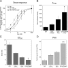
Modulatory activity of TIF2 and STAMP with GR-regulated gene induction of synthetic reporter gene. CV-1 cells were transiently transfected as described in Materials and Methods with GR (6 ng) with or without TIF2 (20 ng) and/or Flag/STAMP (160 ng) plus GREtkLUC reporter (100 ng) and Renilla control (10 ng). After incubation with the indicated concentrations of steroid, vehicle (EtOH), or 1 μM DM in triplicate, the amount of induced luciferase was determined and the data plotted as described in Materials and Methods. (A) Dose-response curves for luciferase induction by GR plus indicated cofactors. Graph is one representative experiment with the error bars being the S.D. of each triplicate steroid treatment. (B) Maximal activity (Amax), (C) steroid potency (EC50) and (D) partial agonist activity (PAA). The average Amax with 1 μM Dex, the average EC50 (nM) with Dex, and the average partial agonist activity of 1 μM DM from four experiments such as in A were determined for the indicated grouping of factors. Error bars equal S.E.M. (n=4). P values for GR+STAMP+TIF2 vs. GR+TIF2 are * = <0.05, ** = < 0.005.
Fig. 2
Fig. 2
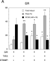
GR-induction properties of endogenous IP6K3 gene with added TIF2 and STAMP in 293 cells. (A) Modulation of parameters of IP6K3 gene induction by transiently transfected full-length GR with TIF2 and STAMP. Experiments were conducted as described in Materials and Methods, with IP6K3 induction being determined by qRT-PCR. Data were then processed and analyzed as in Fig. 1 except that fold induction by 1 μM Dex was quantitated as opposed to Amax. Of relevance is the fact that the basal levels of IP6K3 mRNA expression were essentially the same for each treatment. Note that the y-axis is dimensionless in panels A and C because the bars being plotted have their own associated units (fold induction, percent of maximal activity for PAA, and nM × 10 for EC50). (B) Binding of GR, TIF2, and STAMP to IP6K3 gene. ChIP assays were performed as outlined in Materials and Methods with U2OS cells that were transiently transfected with full-length GR, TIF2, and STAMP followed by treatment with vehicle (EtOH), 1 μM Dex, or 1 μM DM. Primers for PCR amplification were selected to probe protein recruitment to control regions about 2.3 kb upstream and downstream of the transcription start site (TSS), the TSS, a possible pause site at +60-220, and the intronic GRE. Anti-Flag antibody was used to assess non-specific binding of GR, TIF2, and STAMP to each region. Error bars represent S.E.M. (n = 4-6). (C) Induction parameters of IP6K3 gene with truncated GR407C with TIF2 and STAMP. Experiment is the same as in panel A except truncated GR407C is used in place of full-length GR. For both panels A and C, error bars equal S.E.M. (n=4) with P values being for comparisons with data for receptor (GR or GR407C) alone are * = <0.05, ** = < 0.005, *** = <0.0005.
Fig. 2
Fig. 2
GR-induction properties of endogenous IP6K3 gene with added TIF2 and STAMP in 293 cells. (A) Modulation of parameters of IP6K3 gene induction by transiently transfected full-length GR with TIF2 and STAMP. Experiments were conducted as described in Materials and Methods, with IP6K3 induction being determined by qRT-PCR. Data were then processed and analyzed as in Fig. 1 except that fold induction by 1 μM Dex was quantitated as opposed to Amax. Of relevance is the fact that the basal levels of IP6K3 mRNA expression were essentially the same for each treatment. Note that the y-axis is dimensionless in panels A and C because the bars being plotted have their own associated units (fold induction, percent of maximal activity for PAA, and nM × 10 for EC50). (B) Binding of GR, TIF2, and STAMP to IP6K3 gene. ChIP assays were performed as outlined in Materials and Methods with U2OS cells that were transiently transfected with full-length GR, TIF2, and STAMP followed by treatment with vehicle (EtOH), 1 μM Dex, or 1 μM DM. Primers for PCR amplification were selected to probe protein recruitment to control regions about 2.3 kb upstream and downstream of the transcription start site (TSS), the TSS, a possible pause site at +60-220, and the intronic GRE. Anti-Flag antibody was used to assess non-specific binding of GR, TIF2, and STAMP to each region. Error bars represent S.E.M. (n = 4-6). (C) Induction parameters of IP6K3 gene with truncated GR407C with TIF2 and STAMP. Experiment is the same as in panel A except truncated GR407C is used in place of full-length GR. For both panels A and C, error bars equal S.E.M. (n=4) with P values being for comparisons with data for receptor (GR or GR407C) alone are * = <0.05, ** = < 0.005, *** = <0.0005.
Fig. 3
Fig. 3
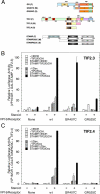
Interaction of GR, TIF2, and STAMP in mammalian three-hybrid assays. (A) Cartoon of different size constructs of each factor. Maps of full (F), medium (M), and short (S) length fragments of GR, TIF2, and STAMP are shown along with the known domains of GR and TIF2. For STAMP, the domains are tyrosine tubulin ligase (TTL), coactivator interaction domain (CID), receptor interaction domain (RID) (He and Simons; Jr., 2007). The full names of the medium and short species of GR and STAMP include a number and the letter “C”, which indicate the first amino acid and the C-terminal amino acid of each species. The TIF2 names are those of Voegel et al. (Voegel et al., 1998). Three-hybrid assays with (B) TIF2.3 or (C) TIF2.4 cotransfected with the indicated VP16/GR fusions, GAL/STAMP623C, and the GAL regulated reporter FRLUC without or with 1 μM Dex. Luciferase activity (in triplicate) was determined as described in Materials and Methods, after which the data were normalized to the highest activity. Error bars in graphs are S.E.M. (n= 2-3 [TIF2.3] or 2 [TIF2.4] independent experiments).
Fig. 4
Fig. 4
Existence of ternary complexes of GR, TIF2, and STAMP. IP/re-IP assays of lightly cross-linked complexes of (A) GR/TIF2/Flag-STAMP and (B) GR524C/TIF2.4/Flag-STAMP623C from cells treated with EtOH (E) or 1 μM Dex (D) were conducted and visualized as described in Materials and Methods. Flag plasmid was used in place of Flag-STAMP as a negative control for the first IP. Anti-TIF2 antibody A is a non-precipitating anti-TIF2 antibody that was used as a negative control for the re-IP. Due to the low levels of expression of Flag-STAMP, it is not detected by Western blotting until after the IP. * = non-specific impurity.
Fig. 5
Fig. 5
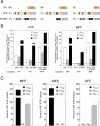
Regions of GR, TIF2, and STAMP required for modulation of Amax, EC50, and PAA. (A) Cartoon of different size constructs of each factor, as described in Fig. 3A. For clarity, the GAL DBD that is fused to GR (S) to give GAL/GR525C is not shown. (B) Effect of factor combinations on induction parameters. The experiments were conducted as in Fig. 1 for different combinations of full-length GR, TIF2, and STAMP (F, F, F) and of medium-length GR with full-length TIF2 and STAMP (M, F, F). The data for each combination of factors are presented as fold increase above that for the GR species of that series by itself for Amax and for 1/EC50 (so that increased potency is displayed as an increase relative to the same GR alone, i.e., GR(M) in MFF series) and as the absolute change in percent agonist activity for PAA (error bars = S.E.M., n = 4 individual experiments). The composition of the binary and ternary complexes in each panel is given at the bottom of each panel. The dashed line represents the level of no change relative to full-length GR. (C) Effect of factor combinations on induction parameters relative to those with full-length proteins. The “percent of wt (FFF) activity” for a given combination (e.g., MFF of panel B) was calculated by dividing the fold increase (or difference in the case of PAA) of the test complex (e.g., MFF) by that of the paired wt standard (e.g., FFF in panel B) as described in the text and Materials and Methods. The data of two other representative series of experiments are also shown for MFS and SFF. NC = no change relative to the paired GR plus TIF2 (or plus STAMP). Neg = negative value compared to the paired GR plus TIF2 (or plus STAMP). It should be noted that the responses of GR + TIF2 are almost always greater than GR + STAMP. Error bars = S.E.M. (n = 4-5).
Fig 6
Fig 6
Activity of modulatory regions of GR, TIF2, and STAMP is cell-dependent. The indicated constructs of GR, TIF2, and STAMP were transiently transfected into 293 cells along with the GREtkLUC reporter. The experiments were conducted and plotted as in Fig. 5. (A) Effect of factor combinations on induction parameters in 293 cells. The data for each combination of factors are presented as in Fig. 5B as fold increase above that for corresponding GR alone for Amax and for 1/EC50 (so that increased potency is displayed as an increase relative to same GR alone, e.g., GR(F) for FMS series) and as the absolute change in percent agonist activity for PAA. The composition of the binary and ternary complexes in each panel is given at the bottom of each panel. The dashed line represents the level of no change relative to full-length GR (error bars = S.E.M., n = 4 individual experiments). (B) Effect of factor combinations on induction parameters relative to those with full-length proteins in 293 cells. The “percent of wt (FFF) activity” for a given combination was calculated from the data in panel A as described for Fig. 5C. NC = no change relative to the paired GR plus TIF2 (or plus STAMP). Neg = negative value compared to the paired GR plus TIF2 (or plus STAMP). Error bars = S.E.M. (n = 4 individual experiments).
Fig. 7
Fig. 7
Model of cofactor modulation of parameters for GR-regulated gene induction. Individual pathways, or interactions with proteins, for altering the levels of gene product can be used such that one, two, or all three of the parameters of Amax, EC50, and PAA are affected. See text for discussion.

Similar articles

Cited by

References

    1. Avenant C, Kotitschke A, Hapgood JP. Glucocorticoid receptor phosphorylation modulates transcription efficacy through GRIP-1 recruitment. Biochemistry. 2010;49:972–985. - PubMed
    1. Barton MC, Shapiro DJ. Transient administration of estradiol-17β establishes an autoregulatory loop permanently inducing estrogen receptor mRNA. Proc. Natl. Acad. Sci. USA. 1988;85:7119–7123. - PMC - PubMed
    1. Biddie SC, John S, Hager GL. Genome-wide mechanisms of nuclear receptor action. Trends Endocrinol Metab. 2010;21:3–9. - PMC - PubMed
    1. Chen W, Rogatsky I, Garabedian MJ. MED14 and MED1 differentially regulate target-specific gene activation by the glucocorticoid receptor. Mol Endocrinol. 2006;20:560–572. - PubMed
    1. Cho S, Blackford JA, Jr., Simons SS., Jr. Role of activation function domain 1, DNA binding, and coactivator in the expression of partial agonist activity of glucocorticoid receptor complexes. Biochemistry. 2005a;44:3547–3561. - PubMed

Publication types

MeSH terms