. 2012 May 15;21(10):2298-311.
doi: 10.1093/hmg/dds050. Epub 2012 Feb 15.

Nxnl2 splicing results in dual functions in neuronal cell survival and maintenance of cell integrity

Affiliations

Nxnl2 splicing results in dual functions in neuronal cell survival and maintenance of cell integrity

Céline Jaillard et al. Hum Mol Genet. .

Abstract

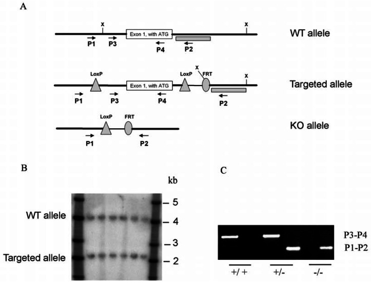
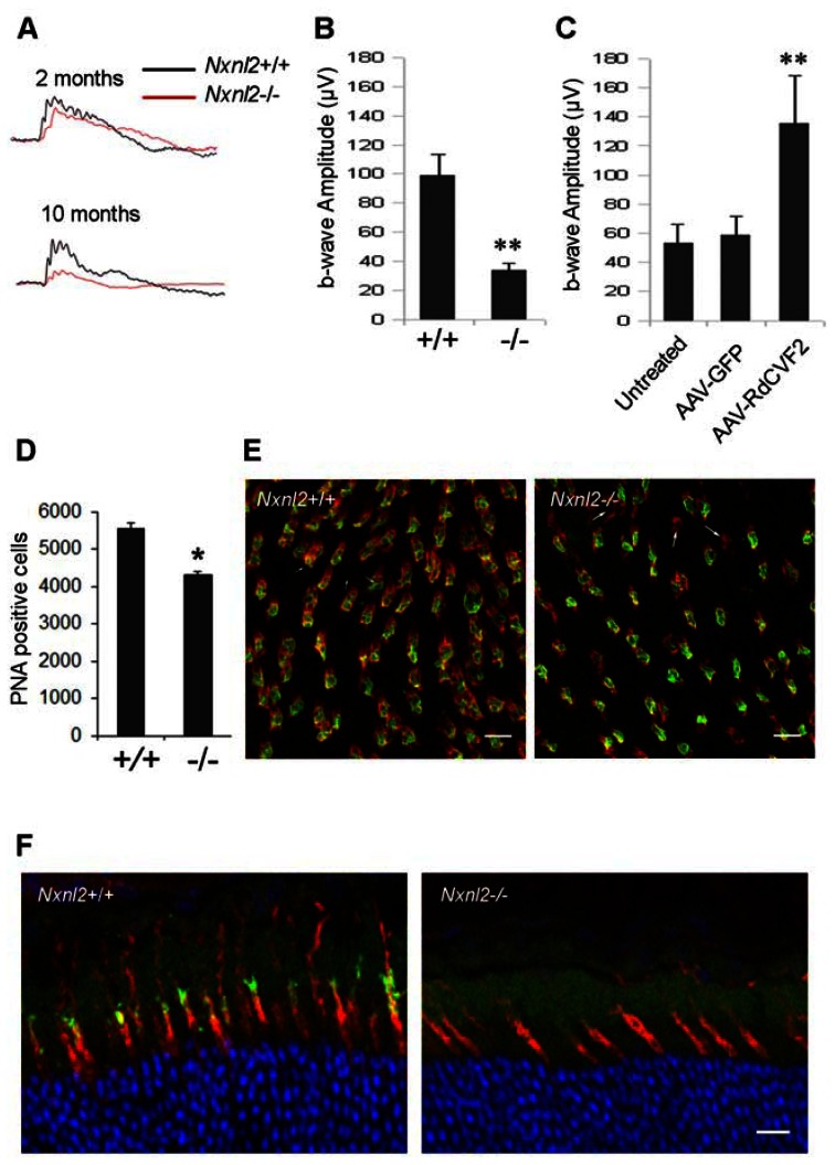
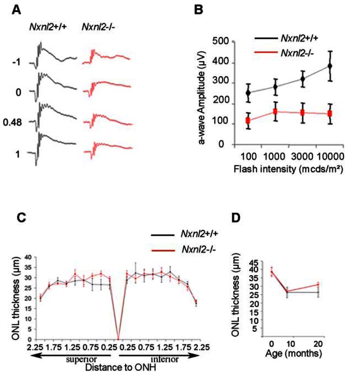
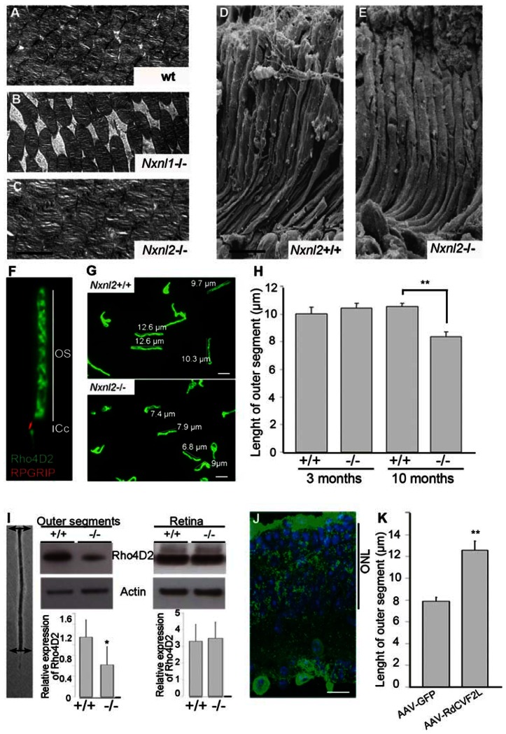
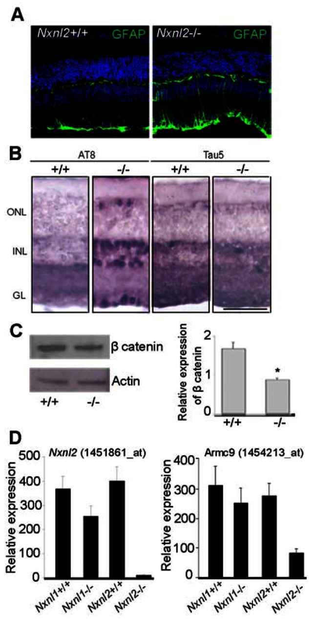
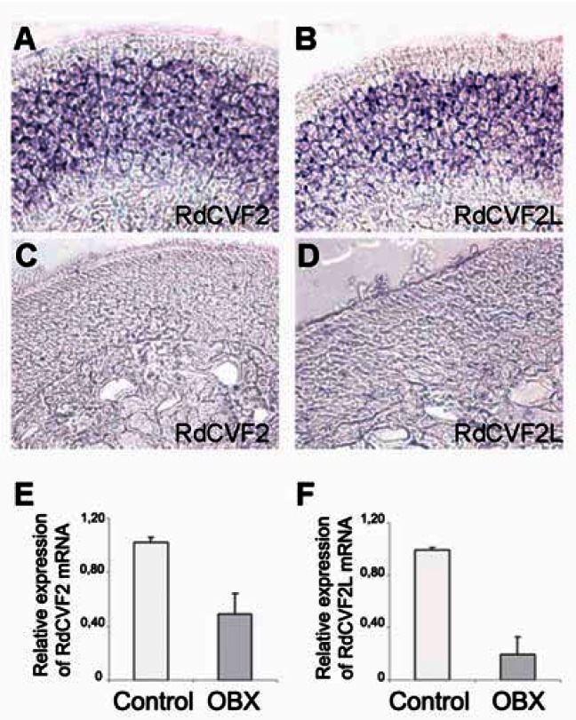
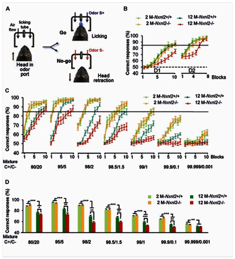
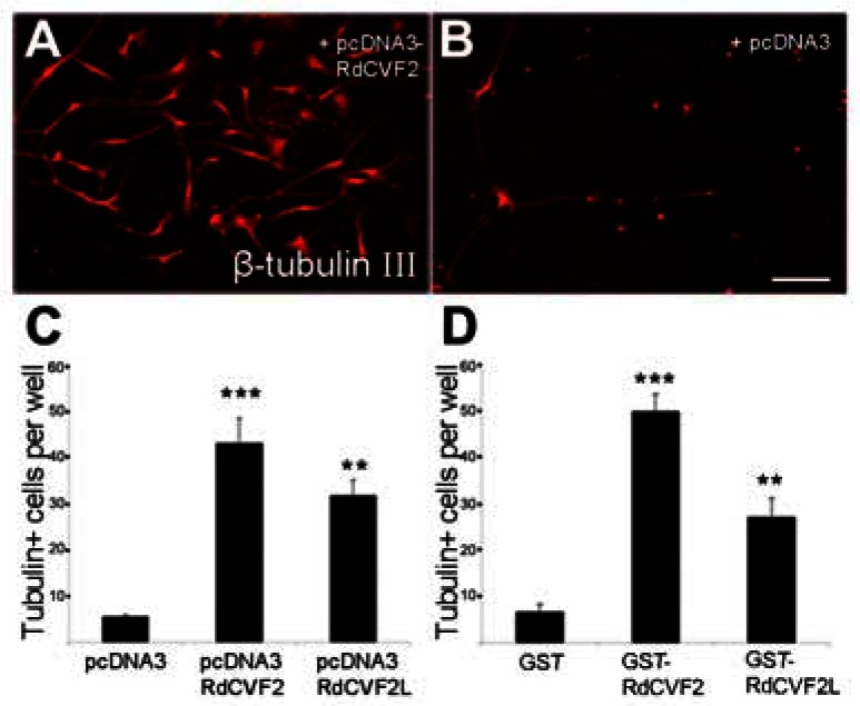
The rod-derived cone viability factors, RdCVF and RdCVF2, have potential therapeutical interests for the treatment of inherited photoreceptor degenerations. In the mouse lacking Nxnl2, the gene encoding RdCVF2, the progressive decline of the visual performance of the cones in parallel with their degeneration, arises due to the loss of trophic support from RdCVF2. In contrary, the progressive loss of rod visual function of the Nxnl2-/- mouse results from a decrease in outer segment length, mediated by a cell autonomous mechanism involving the putative thioredoxin protein RdCVF2L, the second spliced product of the Nxnl2 gene. This novel signaling mechanism extends to olfaction as shown by the progressive impairment of olfaction in aged Nxnl2-/- mice and the protection of olfactory neurons by RdCVF2. This study shows that Nxnl2 is a bi-functional gene involved in the maintenance of both the function and the viability of sensory neurons.


Conflict of interest statement

Conflict of interest statement

CJ, JAS and TL have a patent on Nxnl2 for neurological diseases.

Figures

Figure 1
Figure 1. Nxnl2 targeting strategy
(A) Schema showing the wild-type (WT) Nxnl2 allele, the targeted Nxnl2 locus after homologous recombination and removal of the neomycin selection cassette after Cre mediated loxP recombination and deletion of Nxnl2 exon1. X: XbaI. Dotted box: Southern probe. P1, P2, P3, P4: primer used for PCR genotyping. P1: 5′-TCCTATATGCTGGTTTCCGTC-3′; P2: 5′-TGATCAAGGAGCCTAGCTAAGG-3′; P3: 5′-TCGATTAGAGGTAGAAGAACCC-3′ and P4: 5′-AGCTCCGTGTAGAAGTCGC-3′. (B) Southern hybridization with DNA from ES cells after excision of the neomycin selection cassette. DNA was digested with restriction enzyme XbaI. Hybridization was performed with a 32-P labelled Nxnl2 probe. (C) PCR analysis was performed on DNA isolated from tail of wild-type, heterozygous and homozygous mice for the exon1 Nxnl2 null allele.
Figure 2
Figure 2. Cone degeneration and dysfunction at 10 months in Nxnl2−/− mice
(A) Photopic ERG tracing from wild-type and Nxnl2−/− at 2 and 10 months of age. (B) Summarized photopic ERG data from 10 months of age mice (n = 10). (C) Photopic ERG from Nxnl2−/− mice retina treated at 6 months of age with AAV-GFP, AAV-RdCVF2 or untreated. AAV-RdCVF2 delivery to the retina prevents the loss of cone function observed at 10 months of age in the animal treated with AAV-GFP. (D) Number of cones was evaluated by counting PNA positive cells at 10 months of age. (E) Immunohistochemistry using PNA labeling and S-Opsin antibody on Nxnl2+/+ and Nxnl2−/− flat mounted retina. In Nxnl2−/− retina, the presence of cones labeled with PNA without expression of S-opsin is indicated with an arrow (Scale bar: 5 μm). (F) Immunohistochemistry using PNA labeling and M-Opsin antibody on Nxnl2+/+ and Nxnl2−/− cross sections in ventral part of retina. Scale bar : 20 μm.
Figure 3
Figure 3. Impaired rod function at 10 months in Nxnl2−/− mice
(A) Scotopic ERG tracing from wild-type and Nxnl2−/− at 10 months of age. (B) Summarized scotopic ERG data from 10 months of age (n = 10). (C) Spidergram showing ONL thickness in Nxnl2−/− and control mice at 8 months of age (n = 6). (D) Thinning of Nxnl2−/− and control ONL mice at 3, 8 and 18 months (n = 6).
Figure 4
Figure 4. RdCVF2 is essential to maintenance of outer segment length
(A–C) Ultrastructure of the outer retina in WT, Nxnl1−/− and Nxnl2−/− mice at 12 months of age. The transmission electron microscopy images show the stacked outer segments of the outer nuclear layer. Scale bar : 2 μm. (D) and (E) Scanning electron microscopy on Nxnl2+/+ and Nxnl2−/− retina at 12 months of age showed outer segment morphology. (F) Photoreceptor sensory cilium (PSC) complex was purified and stained with Rho4d2 and RPGRIP antibodies. (G) Outer segments from Nxnl2−/− and control mice at 10 months of age were stained with Rho4D2 antibody. (H) Length of outer segments was evaluated from Nxnl2−/− and control mice at 3 and 10 months of age (n = 3). Scale bars represent 5 μm. (I) Outer segments from control and Nxnl2−/− mouse retina were dissected using laser capture microdissection. Dissected outer segments and whole retina were immunoblotted with rhodopsin and actin antibodies. Quantification was evaluated using Image J software. (J) Representative fluorescence patterns in cryosections 3 months after subretinal injection with AAV2/8-GFP. ONL, outer nuclear layer. Scale bar represents 20 μm. (K) Length of outer segments of Nxnl2−/− mice treated with AAV2/8RdCVF2L.
Figure 5
Figure 5. Signaling pathways in Nxnl2−/− retina
(A) Immunolabeling of cryosections from control and Nxnl2−/− mice at 18 months of age with Glial Fibrillary Acidic Protein (GFAP, green) antibody. (B) Distribution of phosphorylated TAU (AT8) and TAU (Tau5) in 10 months mouse retina. Immunostaining of control and Nxnl2−/− was carried out using AT8 and Tau5 antibodies. GC, ganglion cells; INL, inner nuclear layer; ONL, outer nuclear layer. (C) Western blotting on retinal lysates from control and Nxnl2−/− mice using beta catenin antibody. (D) Relative expression based on the microarray data of a selection of the target genes identified by false discovery rate method in the Nxnl1+/+, Nxnl1−/− Nxnl2+/+ and Nxnl2−/− retina. Scale bar represents 50 μm.
Figure 6
Figure 6. Expression of both Nxnl2 mRNAs by olfactory neurons
(A, B) In situ hybridization on olfactory epithelium sections with a digoxigenin-labeled RdCVF2 and RdCVF2L antisens riboprobes. The specificity of staining was shown with the sense probes (C, D). (E) Quantification of RdCVF2 and RdCVF2L mRNA 6 days post bulbectomy (OBX).
Figure 7
Figure 7. Olfactory discrimination performances of Nxnl2−/− mice decrease with age
(A) Go/No-go procedure in the olfactometer. (B) Mean percentage of correct responses in each block of two days (D1, D2) of training (n = 7–10 mice/group). S+ was (+)-Carvone and S− was (−)-Carvone. 50% represents chance level (dashed line) and 85% represents performance criterion (dotted line). (C) Mean percentage of correct responses for each training block of the seven days (D1 to D7) of training. Mice were trained to discriminate between (+)-Carvone (S+) and a mixture of (+)-Carvone and (−)-Carvone (S−). The concentration of (−)-Carvone in the S− mixture was reduced each day from 2×10⁻⁴ to 5×10⁻⁵, 2×10⁻⁵, 1.5×10⁻⁵, 10−5, 10−6 and 10−8M. Thus, (−)-Carvone successively represented 20%, 5%, 2%, 1.5%, 1%, 0.1% and 0.001% of the mixture. (D) Mean percentage of correct responses for all training blocks for each day of the seven days (D1 to D7). *p < 0.005, **p = 0.01, ***p < 0.001 (n = 7–10). Error bars indicate the SEM. 2 M-WT: wild-type two-month-old mice (n = 9), 2 M-KO: Nxnl2−/− two-month-old mice (n = 9), 12 M-WT: wild-type twelve-month-old mice (n = 10), 12 M-KO: Nxnl2−/− twelve-month-old mice (n = 7).
Figure 8
Figure 8. RdCVF2 promotes survival of adult olfactory sensory neurons (OSNs) in vitro
Survival of OSNs in culture by adding respectively media from COS-1 cells transfected with empty vector pcDNA3, pcDNA-RdCVF2, pcDNA-RdCVF2L (A, B, C) and GST, GST-RdCVF2, GST-RdCVF2L (D).

References

    1. Léveillard T, Mohand-Said S, Lorentz O, Hicks D, Fintz AC, Clerin E, Simonutti M, Forster V, Cavusoglu N, Chalmel F, et al. Identification and characterization of rod-derived cone viability factor. Nat Genet. 2004;36:755–759. - PubMed
    1. Léveillard T, Sahel JA. Rod-derived cone viability factor for treating blinding diseases: from clinic to redox signaling. Sci Transl Med. 2010;2:26ps16. - PMC - PubMed
    1. Mohand-Said S, Deudon-Combe A, Hicks D, Simonutti M, Forster V, Fintz AC, Leveillard T, Dreyfus H, Sahel JA. Normal retina releases a diffusible factor stimulating cone survival in the retinal degeneration mouse. Proc Natl Acad Sci U S A. 1998;95:8357–8362. - PMC - PubMed
    1. Yang Y, Mohand-Said S, Danan A, Simonutti M, Fontaine V, Clerin E, Picaud S, Leveillard T, Sahel JA. Functional cone rescue by RdCVF protein in a dominant model of retinitis pigmentosa. Mol Ther. 2009;17:787–795. - PMC - PubMed
    1. Chalmel F, Leveillard T, Jaillard C, Lardenois A, Berdugo N, Morel E, Koehl P, Lambrou G, Holmgren A, Sahel JA, et al. Rod-derived Cone Viability Factor-2 is a novel bifunctional-thioredoxin-like protein with therapeutic potential. BMC Mol Biol. 2007;8:74. - PMC - PubMed

Publication types