. 2012 Apr;49(4):332-45.
doi: 10.1016/j.fgb.2012.02.001. Epub 2012 Feb 11.

Two cation transporters Ena1 and Nha1 cooperatively modulate ion homeostasis, antifungal drug resistance, and virulence of Cryptococcus neoformans via the HOG pathway

Affiliations

Two cation transporters Ena1 and Nha1 cooperatively modulate ion homeostasis, antifungal drug resistance, and virulence of Cryptococcus neoformans via the HOG pathway

Kwang-Woo Jung et al. Fungal Genet Biol. 2012 Apr.

Abstract

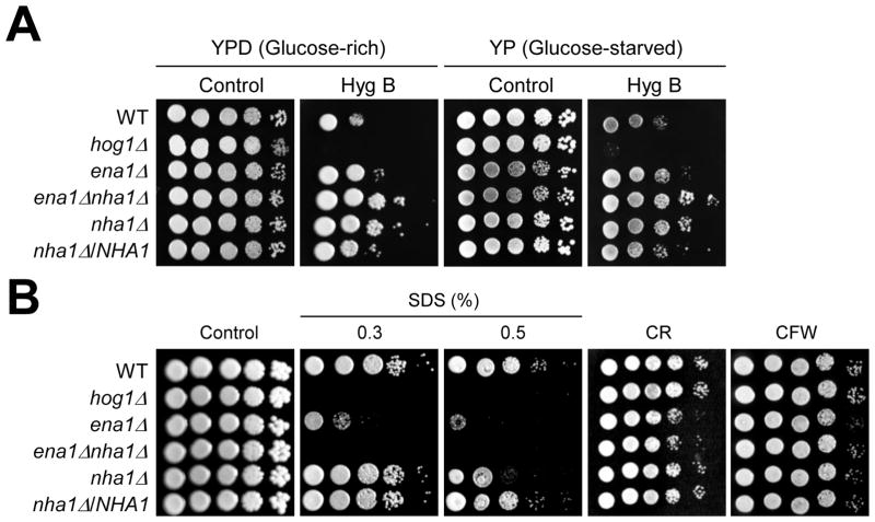
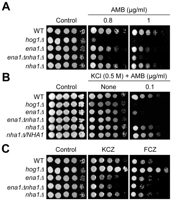
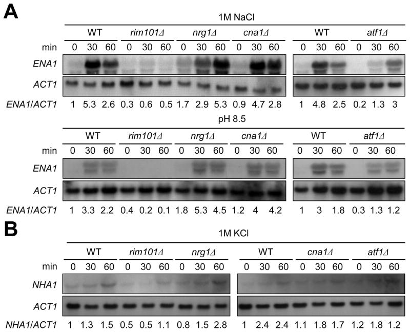
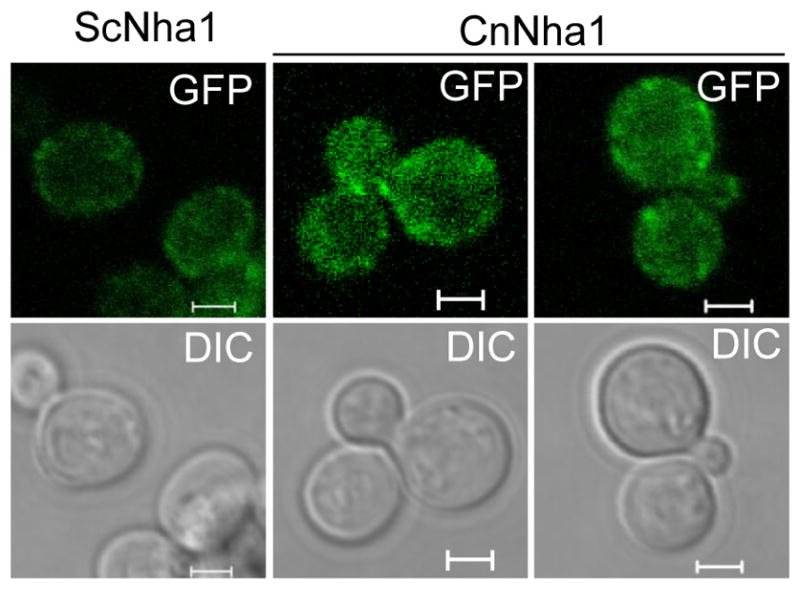
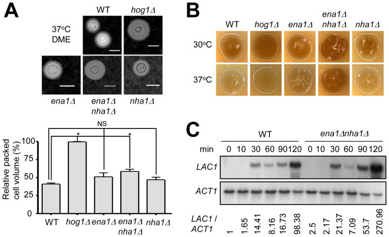
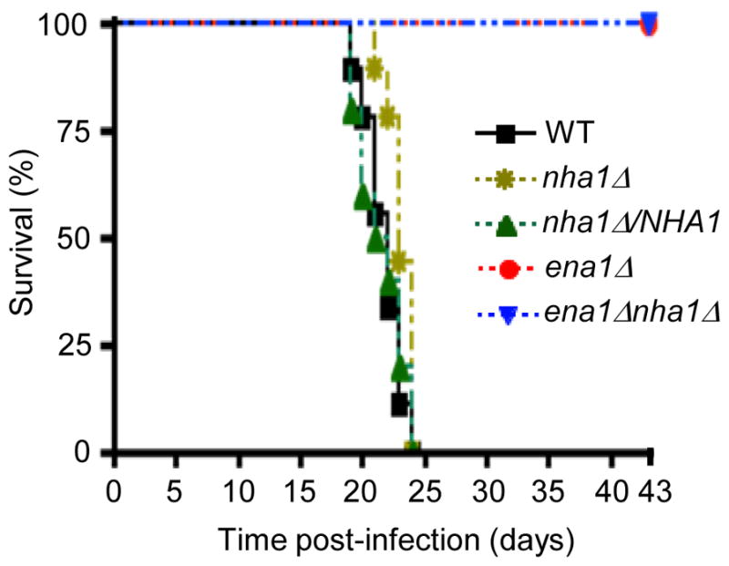
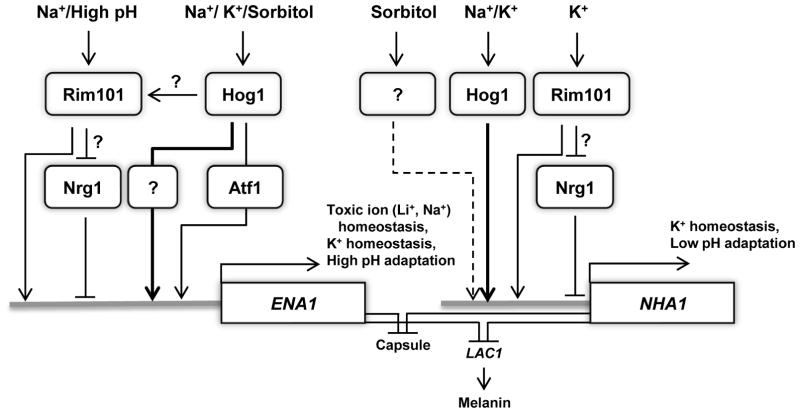
Maintenance of cation homeostasis is essential for survival of all living organisms in their biological niches. It is also important for the survival of human pathogenic fungi in the host, where cation concentrations and pH will vary depending on different anatomical sites. However, the exact role of diverse cation transporters and ion channels in virulence of fungal pathogens remains elusive. In this study we functionally characterized ENA1 and NHA1, encoding a putative Na(+)/ATPase and Na(+)/H(+) antiporter, respectively, in Cryptococcus neoformans, a basidiomycete fungal pathogen which causes fatal meningoencephalitis. Expression of NHA1 and ENA1 is induced in response to salt and osmotic shock mainly in a Hog1-dependent manner. Phenotypic analysis of the ena1Δ, nha1Δ, and ena1Δnha1Δ mutants revealed that Ena1 controls cellular levels of toxic cations, such as Na(+) and Li(+) whereas both Ena1 and Nha1 are important for controlling less toxic K(+) ions. Under alkaline conditions, Ena1 was highly induced and required for growth in the presence of low levels of Na(+) or K(+) salt and Nha1 played a role in survival under K(+) stress. In contrast, Nha1, but not Ena1, was essential for survival at acidic conditions (pH 4.5) under high K(+) stress. In addition, Ena1 and Nha1 were required for maintenance of plasma membrane potential and stability, which appeared to modulate antifungal drug susceptibility. Perturbation of ENA1 and NHA1 enhanced capsule production and melanin synthesis. However, Nha1 was dispensable for virulence of C. neoformans although Ena1 was essential. In conclusion, Ena1 and Nha1 play redundant and discrete roles in cation homeostasis, pH regulation, membrane potential, and virulence in C. neoformans, suggesting that these transporters could be novel antifungal drug targets for treatment of cryptococcosis.


Figures

Fig. 1
Fig. 1
Identification of the NHA1 gene in C. neoformans. (A) The hydrophilicity plot of C. neoformans Nha1 (CnNha1), S. cerevisiae Nha1 (ScNha1), and C. albicans Cnh1 (CaCnh1). The hydrophilicity plots were depicted by protein analysis tool from MacVector software (version 7.2.3, Accelrys). (B) Phylogenetic tree of Nha1 proteins in C. neoformans and other fungi. The phylogenetic tree was generated by philodendron phylogenetic tree printer (http://iubio.bio.indiana.edu/treeapp/treeprint-form.html). The scale bar line represents an evolutionary distance of 0.1.
Fig. 2
Fig. 2
Expression analysis of ENA1 and NHA1 under cation and osmotic stress conditions. Northern blot analysis was performed with total RNA isolated from WTH99 strain and hog1 mutants grown in YPD medium containing 1 M NaCl (A), 1 M KCl (B), or 1 M sorbitol (C) at different time points (0, 10, 30 and 60 min). Each membrane was hybridized with the radioactively labeled ENA1 specific probe, washed, and developed. Subsequently the same membrane was deprobed, re-hybridized with the NHA1 specific probe, washed, and developed.
Fig. 3
Fig. 3
The role of Ena1 and Nha1 in cation homeostasis. (A) WT H99 strain and hog1, ena1, ena1 nha1, and nha1 mutants were grown for 16hr at 30°C in liquid YPD medium, 10-fold serially diluted and spotted on YPD (glucose-rich) or YP (glucose-starved) media containing indicated concentrations of KCl or NaCl. (B) Cells were incubated for 16hr at 30°C in liquid YPD medium, 10-fold serially diluted and spotted on YPD medium containing indicated concentrations of CaCl2 or LiCl.
Fig. 4
Fig. 4
The role of Ena1 and Nha1 in growth under diverse pH conditions. (A) Northern blot analysis for monitoring ENA1 or NHA1 expression in response to different pH conditions. Total RNA was isolated from WT strain H99 grown in different pH conditions as described in Materials and Methods. The relative expression level of ENA1 or NHA1 was quantitatively measured with phosphoimager analysis by normalization with ACT1 expression levels. (B and C) Cells (WT, hog1, ena1, ena1 nha1, nha1 mutants and nha1/NHA1 complemented strain) were spotted on YPD agar medium that was adjusted to acidic (pH 4.5) or alkaline pH (pH 8.5) YPD and contained indicated concentration of KCl or NaCl. (D) Expression levels of NHA1 were monitored under different pH condition (pH 7, pH 4.5, and pH 8.5) in the presence of 1.5 M KCl. The relative expression level of NHA1 was quantitatively measured as described above in (A).
Fig. 5
Fig. 5
The role of Ena1 and Nha1 in maintenance of membrane potential and stability. (A) Cells were spotted onto the YPD or YP medium containing the indicated concentration of Hyg B (Hygromycin B 60 μg/ml and 40 μg/ml in glucose-rich and-starved conditions, respectively). Strains were incubated at 30°C for 3 to 4 days and were photographed. (B) Cells were spotted onto the YPD medium including the indicated concentration of SDS, CR (Congored, 1%) or CFW (Calcofluor white, 2 mg/ml).
Fig. 6
Fig. 6
The role of Ena1 and Nha1 in antifungal drug resistance. (A and C) Strains (WT H99 strain and hog1, ena1, ena1 nha1, and nha1 mutants) were spotted onto YPD medium including the indicated concentration of polyene [AMB (amphotericin B)] or azole [FCZ (fluconazole, 14 μg/ml), KCZ (ketoconazole, 0.3 μg/ml)] drugs. (B) Each C. neoformans strain was grown for 16hr at 30°C in YPD liquid medium, 10-fold serially diluted and spotted YPD (0.5 M KCl) agar containing indicated concentrations of AMB. Cells were incubated at 30°C for 72hr and photographed.
Fig. 7
Fig. 7
Expression patterns of ENA1 and NHA1 in diverse signaling pathways. Total RNA was isolated from WT H99 strain and rim101, nrg1, cna1 and atf1 mutants grown in YPD liquid medium containing 1 M NaCl (A, upper panel) or 1 M KCl (B), or YPD medium adjusted to high pH (8.5) (A, lower panel) at different time points (0, 30, 60 minutes). Each membrane was hybridized with the ENA1 (A) or NHA1 (B) specific probes, washed, and developed. Subsequently the same membrane was deprobed, re-hybridized with the ACT1 specific probe, washed, and developed. Expression levels of ENA1 or NHA1 relative to ACT1 were measured by phosphoimager analysis.
Fig. 8
Fig. 8
Localization of Nha1 in C. neoformans. C. neoformans and S. cerevisiae cells expressing Nha1-Gfpfusion proteins (CnNha1 and ScNha1, respectively) were cultured in SC liquid media at 30°C for 16 hr and were subcultured in SC liquid containing 1 M KCl for 1 h. Cellular localization of Nha1-Gfp proteins were visualized by confocal microscope (Carl Zeiss). The scale bar represents 2 μm.
Fig. 9
Fig. 9
The role of Ena1 and Nha1 in capsule and melanin production. (A) For capsule production measurement, each strain (WT H99 strain and hog1, ena1, ena1 nha1, and nha1 mutants) was spotted and cultured on DME agar medium at 37°C for 2 days. Capsules were visualized by India ink staining (upper panel) and the relative capsule volume was measured by calculating the ratio of the length of packed cell volume phase per length of total volume phase (lower panel). Three independent experiments with technical triplicates were performed. Statistical analysis was performed by using Bonferroni multiple comparison test. *, P < 0.01 and NS, not significant (P > 0.05). (B) Each C. neoformans strain was spotted, cultured on Niger seed medium containing concentration of glucose 0.1% at 30°C or 37°C for 1 day, and photographed. (C) The role of Ena1 and Nha1 in controlling LAC1 expression. WT H99 strain and ena1 nha1 mutants grown to a logarithmic phase (OD600nm = 1.0) in YPD liquid medium (zero time control) were shifted to YNB liquid medium (without glucose), and further incubated at 30°C. Northern blot analysis was performed with total RNA isolated from each cell grown at the indicated time points. Each membrane was hybridized with the LAC1 specific probe, washed, and developed. Subsequently the same membrane was deprobed, re-hybridized with the ACT1 specific probe, washed, and developed. The expression levels of LAC1 relative to ACT1 (LAC1/ACT1) were measured via phosphoimager analysis.
Fig. 10
Fig. 10
Nha1 is not required for virulence in C. neoformans. For virulence assays, seven week old A/J female mice were infected with 5 × 10⁴ cells of WT, ena1, ena1 nha1, nha1, and nha1/NHA1 strains by intranasal inhalation. Survival (%) was monitored daily for 43 days after infection.
Fig. 11
Fig. 11
The proposed model for regulation and function of ENA1 and NHA1 in C. neoformans. Under cation shock and high pH, ENA1 expression is controlled by diverse signaling proteins. ENA1 is positively and negatively regulated by Rim101 and Nrg1 proteins, respectively. However, it is unclear whether Rim101 controls expression of ENA1 through the Nrg1 repressor protein. Hog1 appears to positively regulate ENA1 expression, partly through the Atf1 transcription factor. It is possible that Hog1 controls the Rim101 pathway directly or indirectly to regulate ENA1 expression. Upon Na+/K+ shock, NHA1 expression is induced in a Hog1-dependent manner. Under osmotic shock, such as high sorbitol addition, NHA1 is induced in a Hog1-independent manner. Under K+ shock, Rim101 positively regulated mRNA levels of NHA1. However, it remains unclear whether the Ena1-dependent regulation of NHA1 is mediated via the Nrg1 repressor. In C. neoformans, Ena1 plays a major role in toxic ion (Li+ and Na+) and K+ homeostasis as well as high pH adaptation whereas CnNha1 is involved in K+ homeostasis and low pH adaptation. Furthermore, Ena1 and Nha1 play redundant roles in negatively regulating production of melanin and capsule.

Similar articles

Cited by

References

    1. Alspaugh JA, Perfect JR, Heitman J. Cryptococcus neoformans mating and virulence are regulated by the G-protein α subunit GPA1 and cAMP. Genes Dev. 1997;11:3206–17. - PMC - PubMed
    1. Alspaugh JA, Pukkila-Worley R, Harashima T, Cavallo LM, Funnell D, Cox GM, Perfect JR, Kronstad JW, Heitman J. Adenylyl cyclase functions downstream of the Gα protein Gpa1 and controls mating and pathogenicity of Cryptococcus neoformans. Eukaryot. Cell. 2002;1:75–84. - PMC - PubMed
    1. Arino J. Integrative responses to high pH stress in S. cerevisiae. OMICS. 2010;14:517–23. - PubMed
    1. Bahn YS, Hicks JK, Giles SS, Cox GM, Heitman J. Adenylyl cyclase-associated protein Aca1 regulates virulence and differentiation of Cryptococcus neoformans via the cyclic AMP-protein kinase A cascade. Eukaryot. Cell. 2004;3:1476–91. - PMC - PubMed
    1. Bahn YS, Kojima K, Cox GM, Heitman J. Specialization of the HOG pathway and its impact on differentiation and virulence of Cryptococcus neoformans. Mol. Biol. Cell. 2005;16:2285–300. - PMC - PubMed

Publication types

MeSH terms

LinkOut - more resources