. 2012 May;194(9):2342-54.
doi: 10.1128/JB.06041-11. Epub 2012 Feb 17.

Characterization of Campylobacter jejuni RacRS reveals roles in the heat shock response, motility, and maintenance of cell length homogeneity

Affiliations

Characterization of Campylobacter jejuni RacRS reveals roles in the heat shock response, motility, and maintenance of cell length homogeneity

Dmitry Apel et al. J Bacteriol. 2012 May.

Abstract

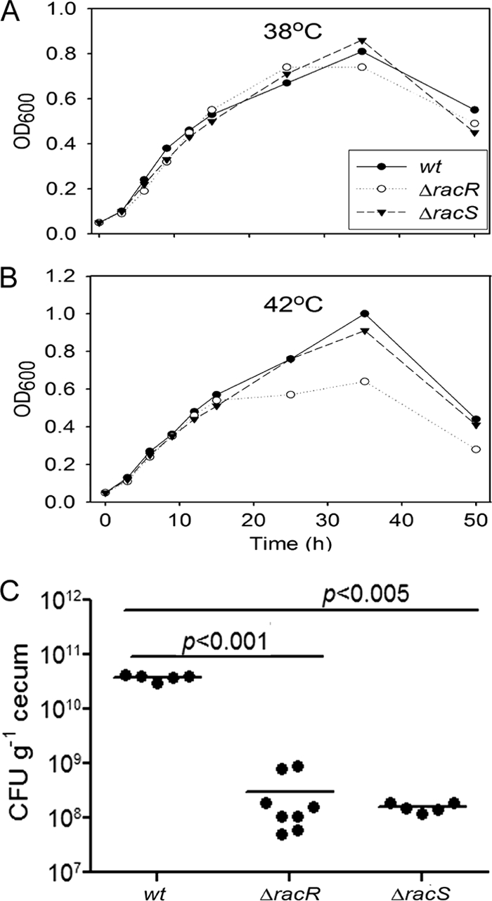
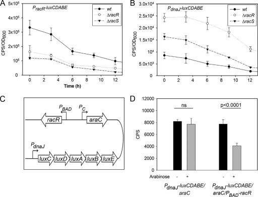
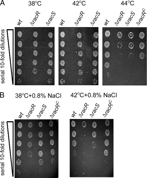
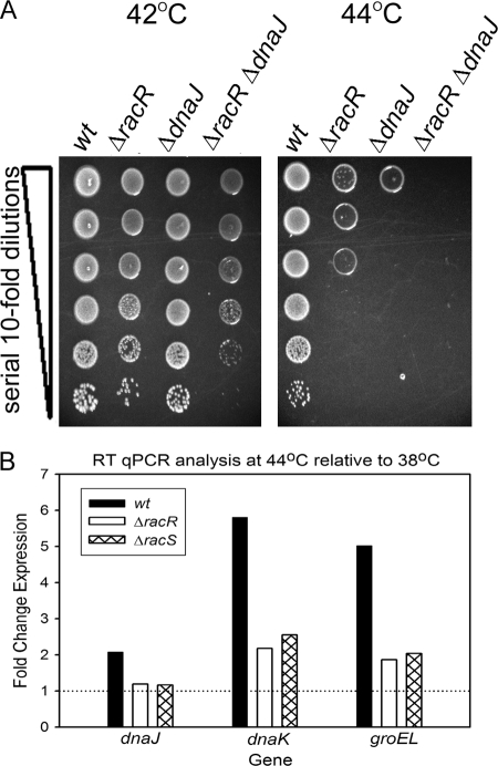
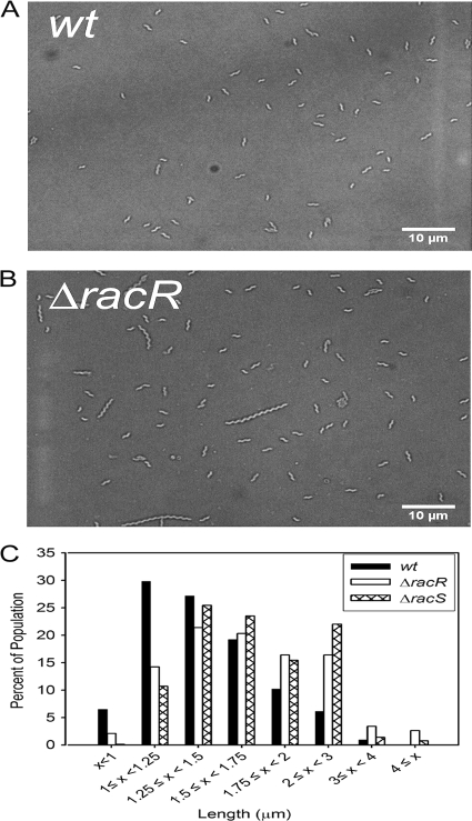
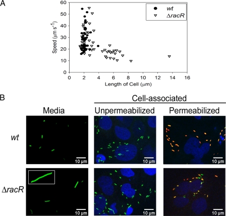
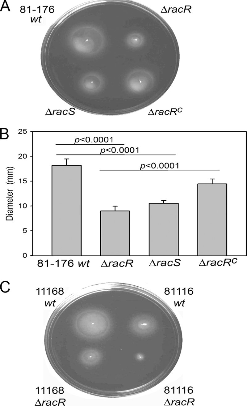
Campylobacter jejuni commensally colonizes the cecum of birds. The RacR (reduced ability to colonize) response regulator was previously shown to be important in avian colonization. To explore the means by which RacR and its cognate sensor kinase RacS may modulate C. jejuni physiology and colonization, ΔracR and ΔracS mutations were constructed in the invasive, virulent strain 81-176, and extensive phenotypic analyses were undertaken. Both the ΔracR and ΔracS mutants exhibited a ~100-fold defect in chick colonization despite no (ΔracS) or minimal (ΔracR) growth defects at 42 °C, the avian body temperature. Each mutant was defective for colony formation at 44°C and in the presence of 0.8% NaCl, both of which are stresses associated with the heat shock response. Promoter-reporter and real-time quantitative PCR (RT-qPCR) analyses revealed that RacR activates racRS and represses dnaJ. Although disregulation of several other heat shock genes was not observed at 38°C, the ΔracR and ΔracS mutants exhibited diminished upregulation of these genes upon a rapid temperature upshift. Furthermore, the ΔracR and ΔracS mutants displayed increased length heterogeneity during exponential growth, with a high proportion of filamented bacteria. Filamented bacteria had reduced swimming speed and were defective for invasion of Caco-2 epithelial cells. Soft-agar studies also revealed that the loss of racR or racS resulted in whole-population motility defects in viscous medium. These findings reveal new roles for RacRS in C. jejuni physiology, each of which is likely important during colonization of the avian host.


Figures

Fig 1
Fig 1
In vitro growth curves of wt and mutant strains at 38°C and 42°C and colonization of day-of-hatch White Leghorn chicks. The growth patterns of 81-176 wt, ΔracR, and ΔracS strains at 38°C (A) and 42°C (B) are shown. Values shown are representative of three independent experiments. OD600, optical density at 600 nm. (C) Chicks were orally inoculated with 104 CFU of C. jejuni resuspended in PBS. At 6 days postinfection, the birds were sacrificed. The bacterial load in the cecum was quantified and is represented as CFU g−1 cecum. Each dot represents the bacterial load in one chicken, and the horizontal lines represent the means.
Fig 2
Fig 2
Measurements of light production of promoter-luciferase fusions in C. jejuni and E. coli. The racR and dnaJ transcriptional regulatory sequences were subcloned in front of the luxCDABE operon in pRY112, and the vector was transformed into wt C. jejuni and the ΔracR and ΔracS mutants. Measurements were made for the first 12 h of bacterial growth. Light counts per second (CPS) were normalized to the OD600 at the time of measurement. (A and B) Data for PracR-luxCDABE (A) and PdnaJ-luxCDABE (B) in wt C. jejuni and the ΔracR and ΔracS mutants are shown. Data are averages of data from 4 independent cultures, and standard deviations are denoted by error bars. (C) Map of the pRY112-derived vector utilized to investigate RacR-dependent transcriptional regulation in E. coli. The luciferase operon was fused to PdnaJ in a plasmid with racR under the transcriptional regulation of the arabinose-inducible PBAD promoter. The transcriptional expression of araC was under regulation of the constitutive promoter (Pc). (D) Light produced by E. coli cultures carrying thePdnaJ-luxCDABE/araC or PdnaJ-luxCDABE/araC/PBAD-racR vector in the presence (+) or absence (−) of arabinose. Error bars denote standard deviations. ns, not significant.
Fig 3
Fig 3
Survival and colony formation of C. jejuni exposed to elevated growth temperatures or osmotic stress. The 81-176 wt, mutant (ΔracR and ΔracS), and complemented (ΔracRC) strains were diluted to an OD600 of 0.1 and spot plated onto solid medium at a serial dilution ranging from 100 to 10−5 (denoted by the vertical wedge). Images were captured after 48 h of incubation at the indicated temperatures on unsupplemented MH plates (A) or on MH plates supplemented with 0.8% NaCl at the indicated temperatures (B) and are representative of three independent experiments.
Fig 4
Fig 4
Survival and colony formation of C. jejuni at elevated growth temperatures and RT-qPCR analysis of C. jejuni exposed to a temperature increase from 38°C to 44°C. (A) wt strain 81-176 and mutant strains (ΔracR, ΔdnaJ, and ΔdnaJ ΔracR) were diluted to an OD600 of 0.1 and spot plated onto solid medium at a serial dilution ranging from 100 to 10−5 (denoted by the vertical wedge). Images were captured after incubation at the indicated temperatures and are representative of three independent experiments. (B) wt C. jejuni 81-176 and mutant strains (ΔracR and ΔracS) were grown to the early log phase in MH broth at 38°C and shifted to 44°C for 15 min. The expression of dnaJ, dnaK, and groEL was analyzed by RT-qPCR. Bars represent the expression ratios from strains shifted to 44°C relative to those that remained at 38°C, with rpoA used as an internal control. The horizontal line indicates no difference in expression levels between the experimental conditions. Data are representative of three independent experiments.
Fig 5
Fig 5
Analysis and quantification of C. jejuni cell length by light microscopy of bacterial populations. Shown are representative images of wt strain 81-176 (A) and the ΔracR strain (B) and the distribution of the length of individual cells of the wt and the ΔracR and ΔracS mutants cultured at 38°C (C). Over 2,000 individual bacteria of each strain from two individual experiments were examined.
Fig 6
Fig 6
Investigation of the impact of C. jejuni cell length on swimming speed and invasion of Caco-2 epithelial cells. (A) The swimming speed of 50 individual wt and ΔracR cells was examined by tracking swimming C. jejuni cells through MH broth by time-lapse microscopy. (B) wt and ΔracR strains expressing GFP were used to infect Caco-2 cells. The bacterium shown in the inset of the ΔracR mutant “Media” panel is ∼30 μm long. To differentiate between extracellular and intracellular bacteria in the cell-associated fractions, those samples were exposed to an anti-C. jejuni antibody either without permeabilization (only extracellular bacteria will be antibody accessible) or following permeabilization with Triton X-100 (extracellular and intracellular bacteria will be antibody accessible) and visualized by using a secondary antibody conjugated to Alexa 568, which appears red. Depending on the relative ratio of GFP expression to antibody reactivity, antibody-accessible bacteria will appear red to yellow-orange. Caco-2 cell nuclei were stained with DAPI and are depicted in blue.
Fig 7
Fig 7
Motility analysis of wt C. jejuni and the ΔracR and ΔracS mutants. (A and B) Representative images of semisolid motility plates incubated for 28 h at 38°C with the 81-176 wt, ΔracR, ΔracS, and ΔracRC strains (A) and quantification of halo diameters (B). Values are averages of data from at least six independent experiments, and the standard deviations are represented by error bars. (C) Representative images of motility plates of wt and ΔracR mutant strains of C. jejuni 11168 and 81116.
Fig 8
Fig 8
Population-level analysis of C. jejuni swimming speed through semisolid medium. (A) The ΔracR mutant bacteria were sampled from either the outer (O), middle (M), or center (C) locations of the migration front of semisolid motility agar. Elongated bacteria were localized at or near the zone of inoculation at the center of the motility halo and are indicated by white arrows. (B) Migration of wt and ΔracR strains through 0.5% and 0.6% agar. (C) wt and ΔracR strains were coinoculated at a 1:1 ratio (wt + ΔracR) in a 0.4% agar motility plate, and the halo diameter was compared to that of either strain inoculated alone. (D) Bacteria from the migration front of a wt- and ΔracR-coinoculated culture were harvested and spot plated onto solid medium with a serial dilution ranging from 100 to 10−5 (denoted by the vertical wedge) on MH medium or MH medium supplemented with kanamycin. In all cases, a representative image is shown.

References

    1. Allen KJ, Griffiths MW. 2001. Effect of environmental and chemotactic stimuli on the activity of the Campylobacter jejuni flaA sigma(28) promoter. FEMS Microbiol. Lett. 205:43–48 - PubMed
    1. Allos BM. 1998. Campylobacter jejuni infection as a cause of the Guillain-Barre syndrome. Infect. Dis. Clin. North. Am. 12:173–184 - PubMed
    1. Allos BM. 2001. Campylobacter jejuni infections: update on emerging issues and trends. Clin. Infect. Dis. 32:1201–1206 - PubMed
    1. Altekruse SF, Stern NJ, Fields PI, Swerdlow DL. 1999. Campylobacter jejuni—an emerging foodborne pathogen. Emerg. Infect. Dis. 5:28–35 - PMC - PubMed
    1. Andersen MT, et al. 2005. Diverse roles for HspR in Campylobacter jejuni revealed by the proteome, transcriptome and phenotypic characterization of an hspR mutant. Microbiology 151:905–915 - PubMed

Publication types

MeSH terms

LinkOut - more resources