. 2012 Apr 4;513(2):160-5.
doi: 10.1016/j.neulet.2012.02.024. Epub 2012 Feb 17.

Diffusion tensor imaging detects axonal injury in a mouse model of repetitive closed-skull traumatic brain injury

Affiliations

Diffusion tensor imaging detects axonal injury in a mouse model of repetitive closed-skull traumatic brain injury

Rachel E Bennett et al. Neurosci Lett. .

Abstract

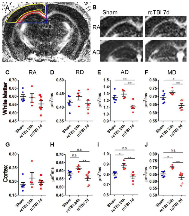
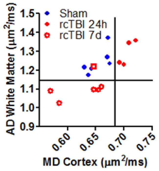
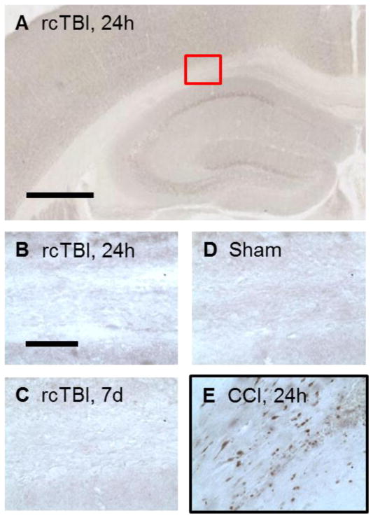
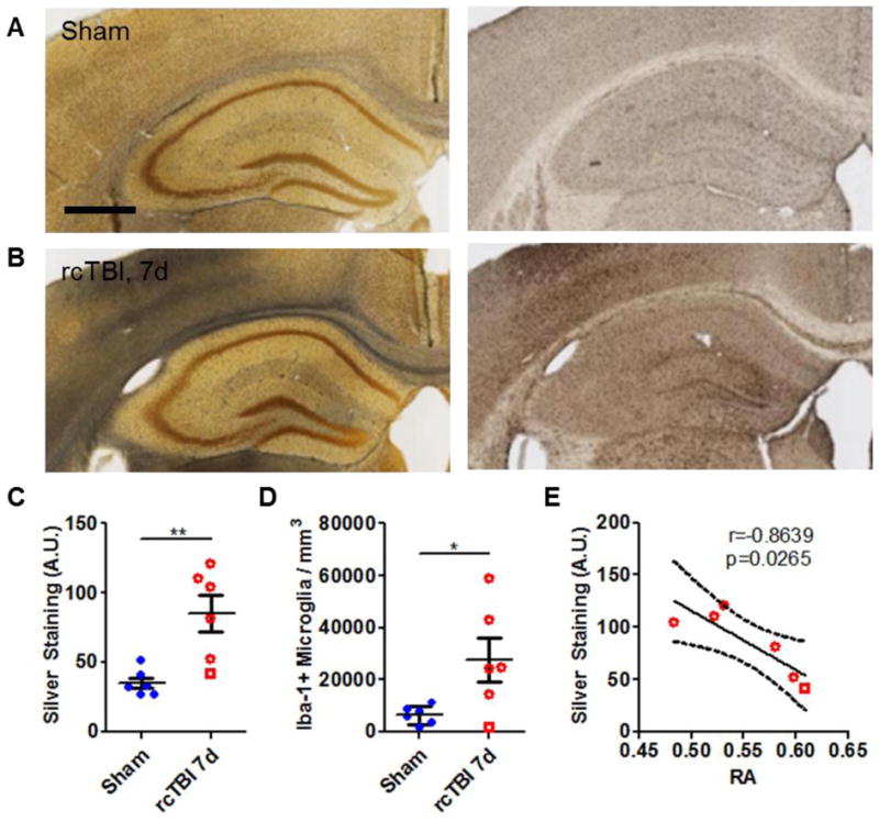
Mild traumatic brain injuries (TBI) are common in athletes, military personnel, and the elderly, and increasing evidence indicates that these injuries have long-term health effects. However, the difficulty in detecting these mild injuries in vivo is a significant impediment to understanding the underlying pathology and treating mild TBI. In the following experiments, we present the results of diffusion tensor imaging (DTI) and histological analysis of a model of mild repetitive closed-skull brain injury in mouse. Histological markers used included silver staining and amyloid precursor protein (APP) immunohistochemistry to detect axonal injury, and Iba-1 immunohistochemistry to assess microglial activation. At 24h post-injury, before silver staining or microglial abnormalities were apparent by histology, no significant changes in any of the DTI parameters were observed within white matter. At 7 days post-injury we observed a reduction in axial and mean diffusivity. Relative anisotropy at 7 days correlated strongly with the degree of silver staining. Interestingly, APP was not observed at any timepoint examined. In addition to the white matter alterations, mean diffusivity was elevated in ipsilateral cortex at 24h but returned to sham levels by 7 days. Altogether, this demonstrates that DTI is a sensitive method for detecting axonal injury despite a lack of conventional APP pathology. Further, this reflects a need to better understand the histological basis for DTI signal changes in mild TBI.


Figures

Figure 1
Figure 1
Diffusion tensor imaging in rcTBI mice. (A) The regions of interest (red, yellow) were defined by anatomical boundaries (blue) and were used for quantitative analysis of diffusion tensor images. (B) Representative images of ipsilateral relative anisotropy (RA) and axial diffusivity (AD) in both a sham and an rcTBI mouse at 7 days. White indicates higher signal intensity and greater RA or AD. (C, G) Relative anisotropy (D, H) Axial diffusivity. (E, I) Radial diffusivity. (F, J) Mean diffusivity. (C–F) White matter. (G–J) Cortex (*p<0.05, **p<0.01, ***p<0.001, one-way ANOVA with Bonferroni post-test, error bars represent ±SEM. Square symbol in 7 day group indicates a mouse with histologically mild injury, a possible outlier.)
Figure 2
Figure 2
Mean diffusivity in cortex versus axial diffusivity in white matter separates mice into distinct injury groups. (Open Square symbol in 7 day group indicates a mouse with histologically mild injury, a possible outlier.)
Figure 3
Figure 3
APP immunohistochemistry. (A, B) Injury did not result in APP positive axons in the corpus callosum of mice analyzed at 24 hours or (C) 7 days following impacts and were similar to (D) sham mice. (E) A mouse subject to moderate controlled cortical impact (CCI) brain injury and sacrificed at 24 hours served as a positive control for APP staining (red box indicates location of B–E, scale bar in A=500 μm, scale bar in B=50 μm).
Figure 4
Figure 4
Axonal injury and microglial activation in 7 day rcTBI. (A) Sham operated mice had little silver stain (black precipitate, left panel) and few Iba-1-positive cells (right panel) in the corpus callosum (scale bar=500 μm). (B) Silver staining (left panel) and increased numbers of Iba-1-positive cells (right panel) were evident in the corpus callosum of mice 7 days post-injury. Quantification of silver by densitometry (C, arbitrary units A.U.) and of Iba-1 by stereology (D) confirmed observed changes (*p<0.05, **p<0.01, Student’s one-tailed t test). (E) Silver staining strongly correlated with changes in relative anisotropy at 7 days post-injury (Pearson’s correlation, two-tailed, solid line represents linear regression, dotted lines represent 95% confidence interval. Square symbol in 7 day group indicates a mouse with a histologically mild injury, a possible outlier).

Similar articles

Cited by

References

    1. Blumbergs PC, Scott G, Manavis J, Wainwright H, Simpson DA, McLean AJ. Staining of amyloid precursor protein to study axonal damage in mild head injury. Lancet. 1994;344:1055–1056. - PubMed
    1. Cubon VA, Putukian M, Boyer C, Dettwiler A. A diffusion tensor imaging study on the white matter skeleton in individuals with sports-related concussion. J Neurotrauma. 2011;28:189–201. - PMC - PubMed
    1. Geddes JF, Whitwell HL, Graham DI. Traumatic axonal injury: practical issues for diagnosis in medicolegal cases. Neuropathol Appl Neurobiol. 2000;26:105–116. - PubMed
    1. Henry LC, Tremblay J, Tremblay S, Lee A, Brun C, Lepore N, Theoret H, Ellemberg D, Lassonde M. Acute and chronic changes in diffusivity measures after sports concussion. J Neurotrauma. 2011;28:2049–2059. - PubMed
    1. Klawiter EC, Schmidt RE, Trinkaus K, Liang HF, Budde MD, Naismith RT, Song SK, Cross AH, Benzinger TL. Radial diffusivity predicts demyelination in ex vivo multiple sclerosis spinal cords. Neuroimage. 2011;55:1454–1460. - PMC - PubMed

Publication types

MeSH terms

LinkOut - more resources