

Hippocampal atrophy and ventricular enlargement in normal aging, mild cognitive impairment (MCI), and Alzheimer Disease
- PMID: 22343374
- PMCID: PMC3286134
- DOI: 10.1097/WAD.0b013e3182163b62
Hippocampal atrophy and ventricular enlargement in normal aging, mild cognitive impairment (MCI), and Alzheimer Disease
Abstract
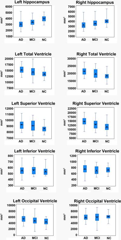
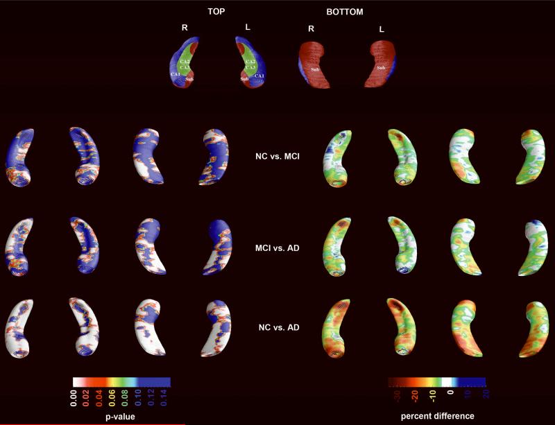
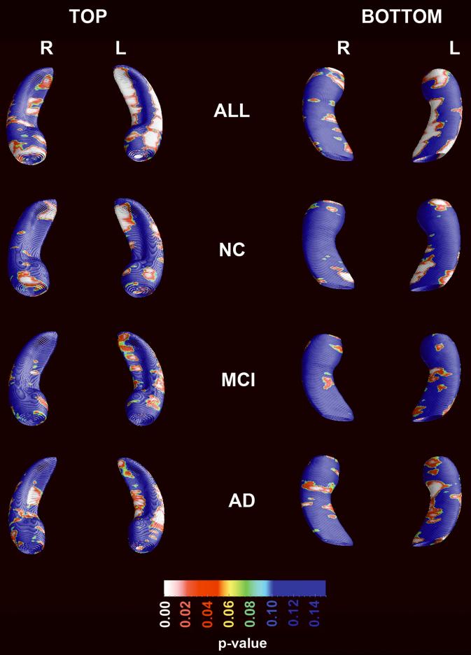
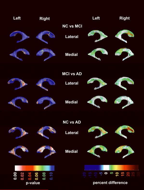
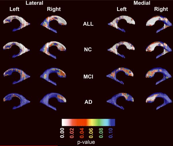
Alzheimer disease (AD) is the most common type of dementia worldwide. Hippocampal atrophy and ventricular enlargement have been associated with AD but also with normal aging. We analyzed 1.5-T brain magnetic resonance imaging data from 46 cognitively normal elderly individuals (NC), 33 mild cognitive impairment and 43 AD patients. Hippocampal and ventricular analyses were conducted with 2 novel semiautomated segmentation approaches followed by the radial distance mapping technique. Multiple linear regression was used to assess the effects of age and diagnosis on hippocampal and ventricular volumes and radial distance. In addition, 3-dimensional map correction for multiple comparisons was made with permutation testing. As expected, most significant hippocampal atrophy and ventricular enlargement were seen in the AD versus NC comparison. Mild cognitive impairment patients showed intermediate levels of hippocampal atrophy and ventricular enlargement. Significant effects of age on hippocampal volume and radial distance were seen in the pooled sample and in the NC and AD groups considered separately. Age-associated differences were detected in all hippocampal subfields and in the frontal and body/occipital horn portions of the lateral ventricles. Aging affects both the hippocampus and lateral ventricles independent of AD pathology, and should be included as covariate in all structural, hippocampal, and ventricular analyses when possible.
(C) 2012 by Lippincott Williams & Wilkins, Inc.
Figures
References
-
- Hebert LE, Scherr PA, Bienias JL, et al. Alzheimer disease in the US population: prevalence estimates using the 2000 census. Archives of Neurology. 2003;60:1119–1122. - PubMed
-
- Wimo A, Jonsson L, Winblad B. An estimate of the worldwide prevalence and direct costs of dementia in 2003. Dement Geriatr Cogn Disord. 2006;21:175–181. - PubMed
-
- Thompson PM, Hayashi KM, De Zubicaray GI, et al. Mapping hippocampal and ventricular change in Alzheimer disease. Neuroimage. 2004;22:1754–1766. - PubMed
Publication types
MeSH terms
Grants and funding
- EB007813/EB/NIBIB NIH HHS/United States
- R01 EB007813/EB/NIBIB NIH HHS/United States
- P50 AG16570/AG/NIA NIH HHS/United States
- HD050735/HD/NICHD NIH HHS/United States
- RC2 AG036535/AG/NIA NIH HHS/United States
- EB008432/EB/NIBIB NIH HHS/United States
- P50 AG016570/AG/NIA NIH HHS/United States
- P41 RR013642/RR/NCRR NIH HHS/United States
- U54 RR021813/RR/NCRR NIH HHS/United States
- EB008281/EB/NIBIB NIH HHS/United States
- R01 MH071940/MH/NIMH NIH HHS/United States
- R01 EB008281/EB/NIBIB NIH HHS/United States
- AG036535/AG/NIA NIH HHS/United States
- K23 AG026803/AG/NIA NIH HHS/United States
- R01 HD050735/HD/NICHD NIH HHS/United States
- R01 EB008432/EB/NIBIB NIH HHS/United States
LinkOut - more resources
Full Text Sources
Medical
Research Materials
