. 2012 May 15;21(10):2341-56.
doi: 10.1093/hmg/dds053. Epub 2012 Feb 17.

Normal myofibrillar development followed by progressive sarcomeric disruption with actin accumulations in a mouse Cfl2 knockout demonstrates requirement of cofilin-2 for muscle maintenance

Affiliations

Normal myofibrillar development followed by progressive sarcomeric disruption with actin accumulations in a mouse Cfl2 knockout demonstrates requirement of cofilin-2 for muscle maintenance

Pankaj B Agrawal et al. Hum Mol Genet. .

Abstract

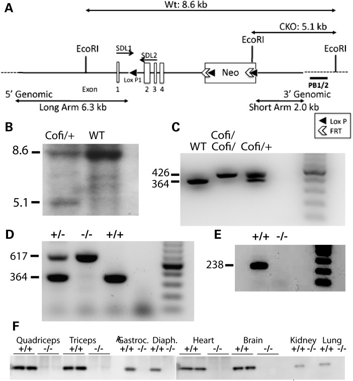
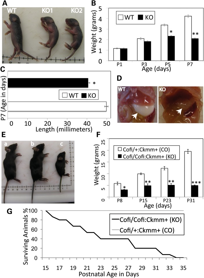
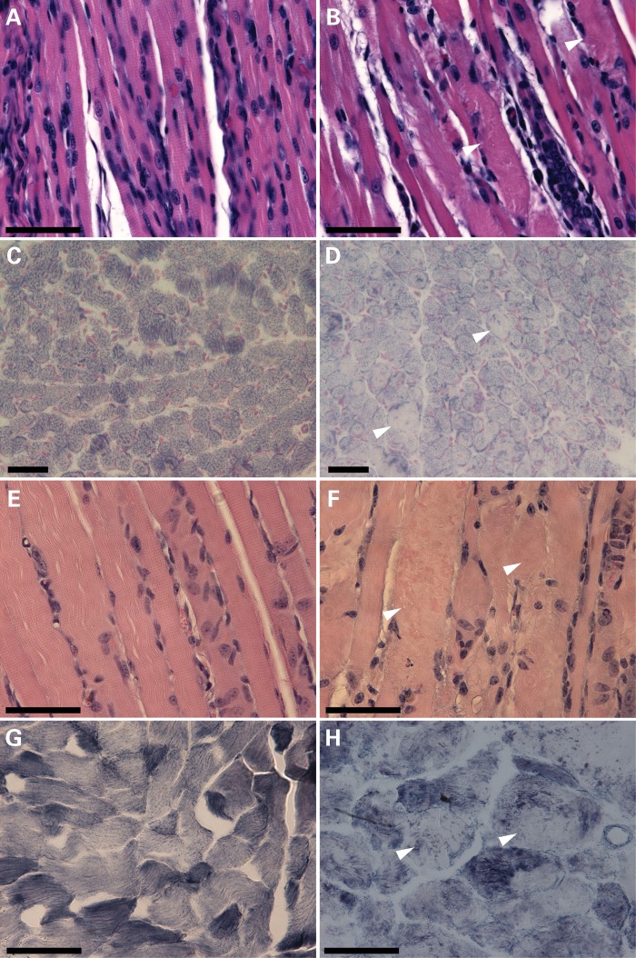
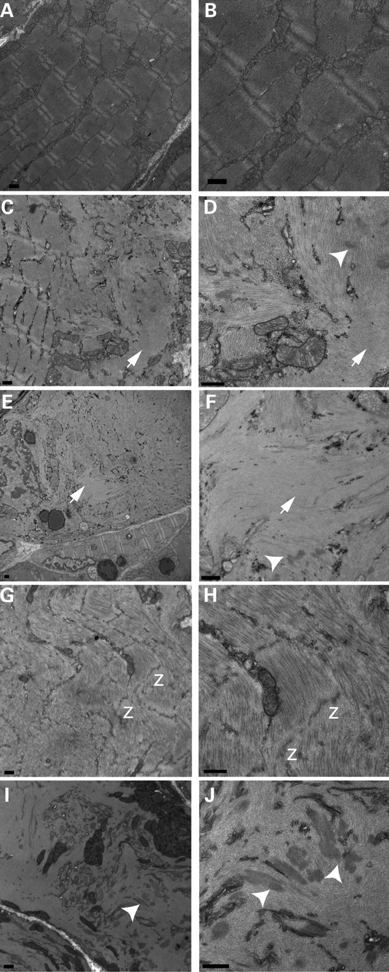
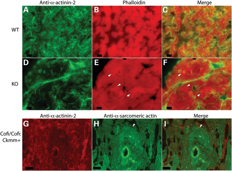
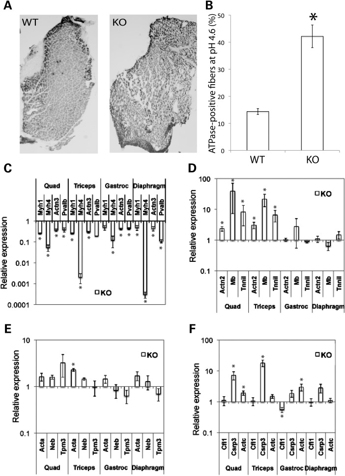
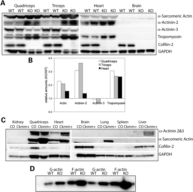
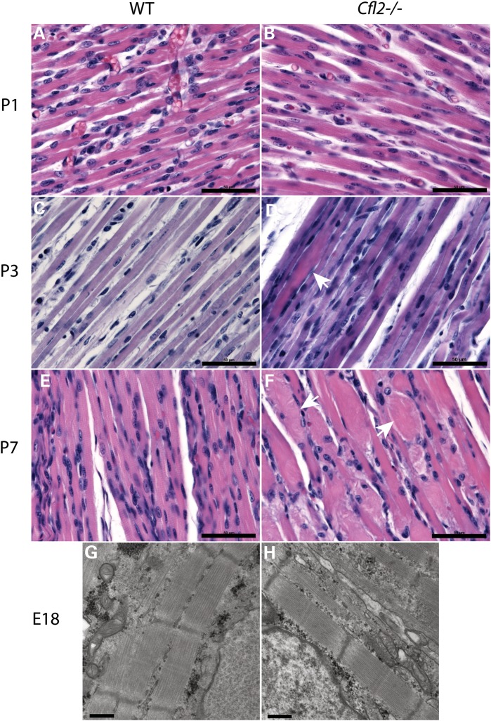
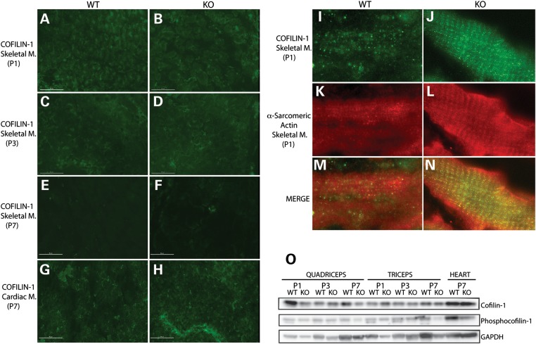
Cofilin-2, a small actin-binding protein and member of the AC protein family that includes cofilin-1 and destrin, is predominantly expressed at sarcomeres in skeletal and cardiac muscles. The role of cofilin-2 in muscle development and function is unclear. In humans, recessive cofilin-2 mutations have been associated with nemaline myopathy with minicores. To investigate the functional role of cofilin-2 in vivo, we generated constitutive and muscle-specific cofilin-2-deficient mice using a cre-loxP strategy. Cofilin-2-deficient mice were similar to their wild-type (WT) littermates at birth, but died by day 8. They were significantly smaller, severely weak and had very little milk in their stomachs. The sarcomeric structure was intact at birth, but by Day 7, skeletal muscles showed severe sarcomeric disruptions starting at the Z-line, along with filamentous actin accumulations consistent with a lack of actin depolymerization activity. Cofilin-2-deficient muscles contained elevated numbers of slow fibers and exhibited upregulation of slow fiber-specific genes. Increased amounts of other sarcomeric proteins including α-actinin-2, α-sarcomeric actin and tropomyosin were also present. While destrin was not expressed in either WT or cofilin-2-deficient muscles, cofilin-1 was similarly expressed in developing myofibers of both genotypes. There was no evidence for compensatory changes in expression of either family member in cofilin-2-deficient tissues. The onset of pathology and weakness in cofilin-2-deficient muscles correlated with normal developmental loss of cofilin-1 expression within myofibers, suggesting that cofilin-1 serves as an early developmental sarcomeric isoform. Overall, cofilin-2, although not critical for muscle development, is essential for muscle maintenance.


Figures

Figure 1.
Figure 1.
Targeting of the Cfl2 gene. (A) Targeting strategy to create Cofi (conditionally targeted Cfl2) allele. An ∼10.4 kb region used to construct the targeting vector was subcloned from a positively identified C57BL/6 BAC clone (RP23-422H22) using a homologous recombination-based technique. The BAC was subcloned into an ∼2.4 kb pSP72 (Promega) backbone vector containing an ampicillin selection cassette. A pGK-gb2 loxP/FRT-flanked Neo cassette was inserted into the gene. The region was designed such that the SA extended 2.0 kb to the 3′ end of loxP/FRT-flanked Neo cassette. The LA extended 6.3 kb to the 5′ end of the single loxP site. The single loxP site was inserted upstream of exon 2, and the loxP/FRT-flanked Neo cassette is inserted downstream of exon 4. The target region was ∼2.1 kb containing exons 2–4. (B) Detection of WT and targeted alleles by Southern blot analysis. DNA isolated from electroporated ES cells was digested with EcoRI and analyzed by Southern blot analysis with a probe against 3′ external region (PB1/2) as shown in (A). The 8.6 and 5.1 kb bands represent WT and targeted alleles, respectively. (C) PCR analysis of DNA isolated from tails of P1 mice showing WT, heterozygous (Cofi/+) and homozygous genotypes (Cofi/Cofi) for the conditionally targeted allele. Primers were designed to include loxP1 present in the Cofi allele to differentiate the two based on sizes, with WT being 364 bp and Cofi 426 bp. (D) Three-primer PCR strategy to confirm excision of two to four exons of Cfl2 along with the Neo cassette. The 364 and 617 bp bands represent the WT (+/+) and Cfl2−/− (−/−) alleles, respectively. (E) RT–PCR using primers for exon 2 (238 bp) confirmed that cofilin-2 was successfully removed in various tissues including the quadriceps. (E) Detection of cofilin-2 protein by western blot analysis. Protein extracted from various tissues of WT (+/+) and Cfl2−/− (−/−) on P7 showed absent cofilin-2 in the quadriceps, triceps, gastrocnemius, diaphragm, heart, brain, kidney and lungs of the Cfl2−/− mice.
Figure 2.
Figure 2.
Phenotypic characterization of cofilin-2-deficient mice. (AD) Cfl2−/− (constitutive cofilin-2-KO) mice. (A) The Cfl2−/− mice (KO1 and KO2) were significantly smaller than a WT littermate. (B) The mean weights for KO (Cfl2−/−) and WT mice were similar at birth (P1) and started to trend lower on P3, and by P5, KO mice were significantly lighter than the WT controls (P < 0.005). On P7, the KO mice weighed less than half that of the WT (P < 0.0001). (C) The length of KO mice was significantly less than that of WT littermates on P7 (P < 0.0001). (D) The stomach bubble (arrows) in KO mice was empty while WT stomachs were distended with milk. (EG) Cofi/Cofi:Ckmm+ mice (muscle-specific cofilin-2 KO with post-natal excision of Cfl2). (E) The Cofi/Cofi:Ckmm+ mice (c) were significantly smaller compared with the age-matched WT (a) and littermate Cofi/+:Ckmm+ (b) mice as controls. (F) The mean weights of KO mice were significantly lower (Cofi/Cofi:Ckmm+) on P8 (P < 0.05), P15 (P < 0.005), P23 (P < 0.0005) and P31 (P< 0.0001) compared with their Cofi/+:Ckmm+ littermates as controls (CO). (G) The Kaplan–Meier curve showing survival of the Cofi/Cofi:Ckmm+ (KO) mice (median 21 days, range 16–33 days, n = 15).
Figure 3.
Figure 3.
Skeletal muscle histopathology of cofilin-2-deficient mice (Cfl2−/− and Cofi/Cofi:Ckmm+). H&E staining (A, B, E and F) of quadriceps cross-sections from WT (A) and Cfl2−/− (B) at P7, and Cofi/+:Ckmm+ (E) and Cofi/Cofi:Ckmm+ (F) mice at P21 was performed. Sections from Cfl2−/− and Cofi/Cofi:Ckmm+ muscles revealed ballooning sarcomeric degeneration (arrowheads). NADH staining (C, D, G and H) was performed on quadriceps cross-sections from WT (C), Cfl2−/− (D), Cofi/+:Ckmm+ (G) and Cofi/Cofi:Ckmm+ (H) mice. Sections from Cfl2−/− and Cofi/Cofi:Ckmm+ mice revealed pale areas within the myofibers as shown by arrowheads (scale bar = 50 μm).
Figure 4.
Figure 4.
Transmission EM of cofilin-2-deficient skeletal muscles. Samples were obtained from WT (quad, A and B), Cfl2−/− (triceps, C and D), Cofi/Cofi:Acta1+ (quad, E and F), Cofi/Cofi:Mef2c+ (quad, G and H) and Cofi/Cofi:Ckmm+ (triceps, I and J) mice. Sarcomeric disruptions were present in fibers from cofilin-2-deficient mice (C–J). WT fibers showing intact sarcomeres for comparison (A and B). Sarcomeric disruptions included intact sarcomeres alongside disrupted ones within a fiber (C) or an intact fiber alongside a disrupted fiber (E). Actin accumulations shown by arrows (C, D, G and H) and nemaline bodies shown by arrowheads (D, F, I and J) were present within disrupted fibers. Disintegrating Z-lines were noted, shown by ‘z’ (G and H) (scale bar = 500 nm).
Figure 5.
Figure 5.
Fluorescence microscopic analysis revealing actin filament accumulations. Muscle sections from WT (AC), KO (Cfl2−/−) (DF) and Cofi/Cofi:Ckmm+ (GI) mice were immunostained with anti-α-actinin-2 (A, D and G), phalloidin (B and E) and anti-α-sarcomeric actin (H) antibodies. Merged images are shown in (C), (F) and (I). Several actin accumulations are indicated by arrowheads in (E), (F), (H) and (I) (scale bar = 10 µm).
Figure 6.
Figure 6.
Fiber-type distribution in triceps of WT and Cfl2−/− mice on P7 and expression analysis of fiber-type-specific genes. Frozen sections of triceps from KO (Cfl2−/−) and WT littermates at P7 assayed for myosin ATPase activity (pH 4.6), demonstrating relative proportions of slow (dark stained) and fast (light stained) fibers. (A) KO triceps had more darkly stained fibers on microscopy. (B) Slow fibers and total number of fibers were counted for sections taken from the triceps (n = 4). The percentage of slow fibers in the WT and KO triceps was calculated, and the mean difference was found to be statistically significant (P < 0.05). Expression of fast fiber-specific genes (C) and slow fiber-specific genes (D) in various muscle groups from WT and KO mice (n = 4) was measured using qRT–PCR and analyzed. Expression of genes encoding thin filament proteins (E) and expression of Cfl1, Csrp3 and Actc (F) were also compared. Relative expression was compared using mean fold change (KO/WT) ± standard error, and P-values using t-test were calculated (*P < 0.05).
Figure 7.
Figure 7.
Western blot analysis of sarcomeric protein expression in skeletal muscles, the heart and other tissues from Cfl2−/− and Cofi/Cofi:Ckmm+ mice. (A) Immunoblot analysis of P7 KO (Cfl2−/−) and WT littermate in duplicate. Increased levels of α-sarcomeric actin, α-actinin-2 and sarcomeric tropomyosin were present in the KO quadriceps, triceps and the heart. Decreased levels of α-actinin-3 were present in the KO quadriceps and triceps (absent in the heart). Cofilin-2 was expressed in the brain, but α-sarcomeric actin, α-actinin-2, α-actinin-3 and tropomyosin were absent. (B) Densitometric analysis using Quantity One software confirmed the increase in sarcomeric proteins as described above. (C) Immunoblot analysis of tissues from 3-week-old Cofi/Cofi:Ckmm+ (Ckmm+) and control (CO) littermate. Cofilin-2 levels were markedly reduced in the quadriceps and heart but were similar in other tissues including the kidney, brain, lung, spleen and liver. Increased levels of α-sarcomeric actin in the quadriceps, heart and presence of actin in lungs were noted in the Ckmm+ mice. (D) Assessment of G-actin and F-actin levels in the frozen muscle extracts of WT and KO mice (in duplicate) by separating the two fractions by centrifugation and probing blots using antibody against α-sarcomeric actin. Both G-actin and F-actin were increased in the KO, but an increase in F-actin was relatively higher.
Figure 8.
Figure 8.
Evolution of muscle degeneration in Cfl2−/− mice (B, D, F and H) and comparison with WT littermate (A, C, E and G). On P1, muscles from the WT and Cfl2−/− mice were very similar on H&E staining (A and B). By P3, occasional degenerating fibers were noted (arrow) in Cfl2−/− mice (D). On P7, multiple degenerated fibers (arrows) were present in the Cfl2−/− mice muscles (F) (scale bar = 50 µm). EM on a section from E18 embryos showed normally developed myofibrils in both WT and Cfl2−/− mice (scale bar = 500 nm).
Figure 9.
Figure 9.
Cofilin-1 expression in WT and KO (Cfl2−/−) mice. Immunofluorescence analysis to evaluate cofilin-1 expression in skeletal muscle samples obtained from WT and KO mice on P1 (A and B), P3 (C and D) and P7 (E and F), and cardiac muscle samples on P7 (G and H) using anti-cofilin-1-specific antibody. In skeletal muscles, progressive reduction in cofilin-1 expression was noted with age (A, C and E for WT and B, D and F for KO), and on P7, cofilin-1 was absent from within myofibers in WT (E) and KO mice (F). In the cardiac muscle, cofilin-1 remained present within the myofibers (G and H) on P7 (scale bar = 50 μm). Evaluation of cofilin-1 localization using anti-cofilin-1 (I and J)- and anti-α-sarcomeric actin (K and L)-specific antibodies, along with merged images (M and N), in skeletal muscles from WT and KO mice on P1. Cofilin-1 was present in a striated pattern, and co-localized with α-sarcomeric actin in both WT (I, K and M) and KO (J, L and N) mice. (O) Western blot analysis of cofilin-1 and phosphocofilin-1 levels in the WT and KO quadriceps, triceps and heart on P1, P3 and P7. The amounts of cofilin-1 and phosphocofilin-1 were similar between the WT and KO quadriceps, triceps and heart on P1, P3 and P7.

Similar articles

Cited by

References

    1. Bamburg J.R. Proteins of the ADF/cofilin family: essential regulators of actin dynamics. Annu. Rev. Cell Dev. Biol. 1999;15:185–230. - PubMed
    1. Agrawal P.B., Greenleaf R.S., Tomczak K.K., Lehtokari V.L., Wallgren-Pettersson C., Wallefeld W., Laing N.G., Darras B.T., Maciver S.K., Dormitzer P.R., et al. Nemaline myopathy with minicores caused by mutation of the CFL2 gene encoding the skeletal muscle actin-binding protein, cofilin-2. Am. J. Hum. Genet. 2007;80:162–167. - PMC - PubMed
    1. Thirion C., Stucka R., Mendel B., Gruhler A., Jaksch M., Nowak K.J., Binz N., Laing N.G., Lochmuller H. Characterization of human muscle type cofilin (CFL2) in normal and regenerating muscle. Eur. J. Biochem. 2001;268:3473–3482. - PubMed
    1. Mohri K., Takano-Ohmuro H., Nakashima H., Hayakawa K., Endo T., Hanaoka K., Obinata T. Expression of cofilin isoforms during development of mouse striated muscles. J. Muscle Res. Cell Motil. 2000;21:49–57. - PubMed
    1. Vartiainen M.K., Mustonen T., Mattila P.K., Ojala P.J., Thesleff I., Partanen J., Lappalainen P. The three mouse actin-depolymerizing factor/cofilins evolved to fulfill cell-type-specific requirements for actin dynamics. Mol. Biol. Cell. 2002;13:183–194. - PMC - PubMed

Publication types