. 2012 May 1;52(9):1666-79.
doi: 10.1016/j.freeradbiomed.2012.02.010. Epub 2012 Feb 15.

P2X7 receptor-NADPH oxidase axis mediates protein radical formation and Kupffer cell activation in carbon tetrachloride-mediated steatohepatitis in obese mice

Affiliations

P2X7 receptor-NADPH oxidase axis mediates protein radical formation and Kupffer cell activation in carbon tetrachloride-mediated steatohepatitis in obese mice

Saurabh Chatterjee et al. Free Radic Biol Med. .

Abstract

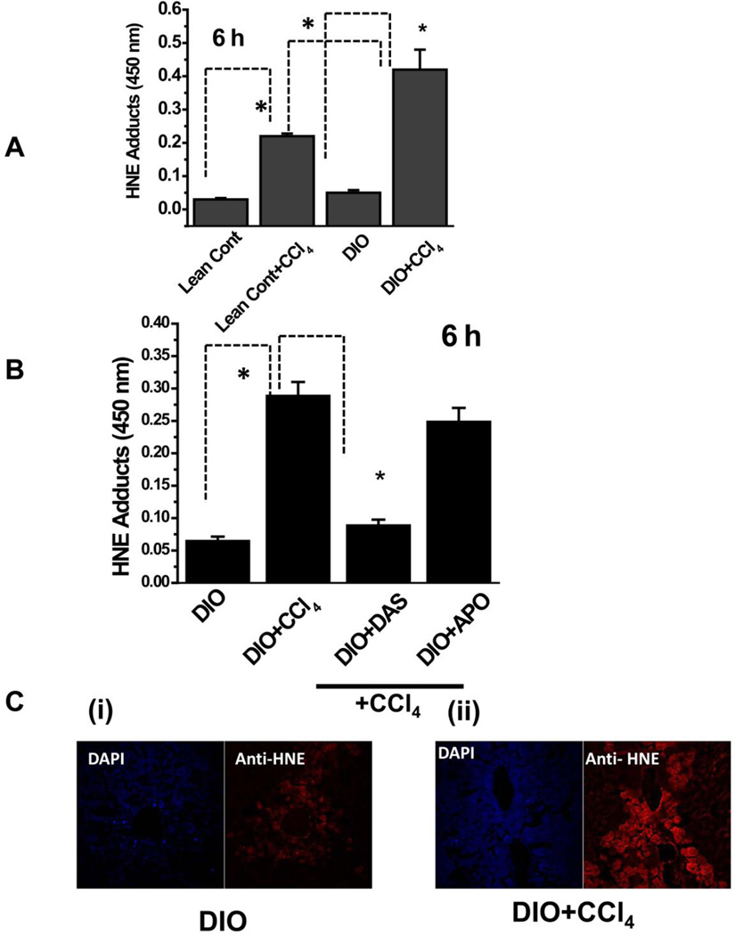
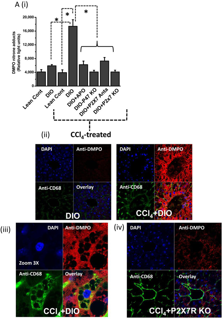
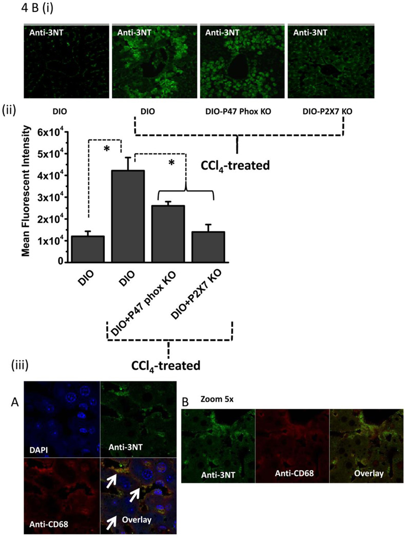
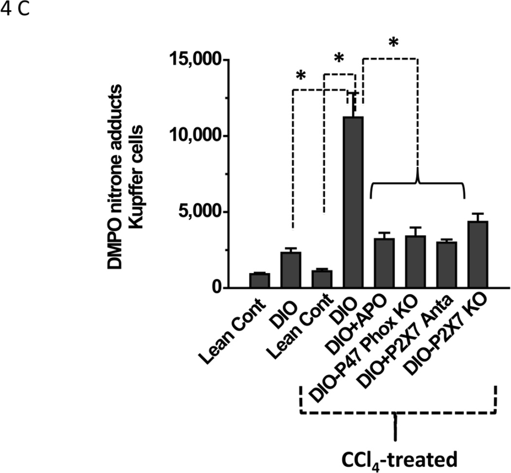
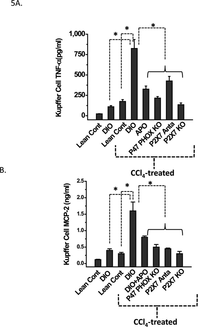
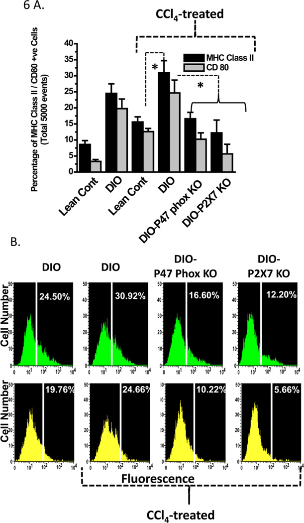
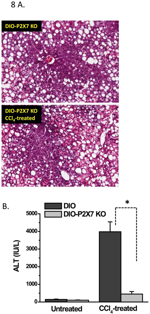
While some studies show that carbon tetrachloride-mediated metabolic oxidative stress exacerbates steatohepatitic-like lesions in obese mice, the redox mechanisms that trigger the innate immune system and accentuate the inflammatory cascade remain unclear. Here we have explored the role of the purinergic receptor P2X7-NADPH oxidase axis as a primary event in recognizing the heightened release of extracellular ATP from CCl(4)-treated hepatocytes and generating redox-mediated Kupffer cell activation in obese mice. We found that an underlying condition of obesity led to the formation of protein radicals and posttranslational nitration, primarily in Kupffer cells, at 24h post-CCl(4) administration. The free radical-mediated oxidation of cellular macromolecules, which was NADPH oxidase and P2X7 receptor-dependent, correlated well with the release of TNF-α and MCP-2 from Kupffer cells. The Kupffer cells in CCl(4)-treated mice exhibited increased expression of MHC Class II proteins and showed an activated phenotype. Increased expression of MHC Class II was inhibited by the NADPH oxidase inhibitor apocynin , P2X7 receptor antagonist A438709 hydrochloride, and genetic deletions of the NADPH oxidase p47 phox subunit or the P2X7 receptor. The P2X7 receptor acted upstream of NADPH oxidase activation by up-regulating the expression of the p47 phox subunit and p47 phox binding to the membrane subunit, gp91 phox. We conclude that the P2X7 receptor is a primary mediator of oxidative stress-induced exacerbation of inflammatory liver injury in obese mice via NADPH oxidase-dependent mechanisms.


Figures

Figure 1
Figure 1
Lipid peroxidation in CCl4-treated DIO mouse liver. A. ELISA of HNE adducts of liver from lean control and DIO mice untreated or treated with CCl4. B. Effect of CYP2E1 inhibitor diallyl sulfide (DAS) or NADPH oxidase inhibitor apocynin (APO) on lipid peroxidation in DIO mice treated with CCl4. C. Confocal micrographs of liver sections of DIO mice (untreated, left panel, and treated with CCl4 ,right panel. * Significantly different (P<0.05).
Figure 2
Figure 2
Metabolic oxidative stress from CCl4 potentiates early steatohepatitic lesions in obesity. A. Eosin-hematoxylin-stained mouse liver sections showing micro and macro vesicular steatosis, portal and lobular necroinflammation and balloonic degeneration at 48 h post CCl4 administration. (i) lean control, (ii) lean control treated with CCl4, (iii) DIO mouse liver and (iv) DIO + CCl4. Blue arrows indicate portal and lobular accumulation of immune cells. Yellow arrow indicates balloon degeneration of hepatocytes. Yellow bordered inlet shows 40x magnified image. B. Representative transmission electron microscopic images of mouse livers treated with CCl4. (i) lean control and (ii) diet-induced obese mice. Significantly degenerating hepatocytes and mitochondria with clumped cristae can be seen in DIO liver section. SER: Smooth endoplasmic reticulum; M: Mitochodria; RER: Rough endoplasmic reticulum; L: Lipid droplets; NT: Neutrophil; EN: Endothelial cell nucleus; GL: Globular lipid; SL: Sinusoidal lumen; LY: Lysosome. C.Serum ALT levels in diet-induced obese mice treated with 120 mg/kg (0.8 mM) CCl4 at 24 h. D. Oil Red O staining of liver sections from Lean control, lean control+CCl4, DIO and DIO+CCl4 groups showing extent of steatosis.
Figure 2
Figure 2
Metabolic oxidative stress from CCl4 potentiates early steatohepatitic lesions in obesity. A. Eosin-hematoxylin-stained mouse liver sections showing micro and macro vesicular steatosis, portal and lobular necroinflammation and balloonic degeneration at 48 h post CCl4 administration. (i) lean control, (ii) lean control treated with CCl4, (iii) DIO mouse liver and (iv) DIO + CCl4. Blue arrows indicate portal and lobular accumulation of immune cells. Yellow arrow indicates balloon degeneration of hepatocytes. Yellow bordered inlet shows 40x magnified image. B. Representative transmission electron microscopic images of mouse livers treated with CCl4. (i) lean control and (ii) diet-induced obese mice. Significantly degenerating hepatocytes and mitochondria with clumped cristae can be seen in DIO liver section. SER: Smooth endoplasmic reticulum; M: Mitochodria; RER: Rough endoplasmic reticulum; L: Lipid droplets; NT: Neutrophil; EN: Endothelial cell nucleus; GL: Globular lipid; SL: Sinusoidal lumen; LY: Lysosome. C.Serum ALT levels in diet-induced obese mice treated with 120 mg/kg (0.8 mM) CCl4 at 24 h. D. Oil Red O staining of liver sections from Lean control, lean control+CCl4, DIO and DIO+CCl4 groups showing extent of steatosis.
Figure 3
Figure 3
Measurement of extracellular ATP, a danger associated molecular pattern and a ligand for purinergic receptors. Extracellular ATP was measured from supernatants of CD-1 mouse primary hepatocytes at different time points co-cultured with 5 mM CCl4 (upper panel) and ATP release from hepatocytes (lower panel) that were isolated from lean and DIO mice. The hepatocytes were co-cultured with 5 mM CCl4 for 24 hours. ATP measurement was done on harvested cells at 24h. * Significantly different (P<0.05).
Figure 4
Figure 4
Metabolic oxidative stress forms protein radicals and post translational tyrosine nitration in Kupffer cells in NADPH oxidase and P2X7 receptor-dependent mechanisms. A. Formation of protein radical adducts in intact liver sections and homogenates using ELISA and confocal microscopy at 24 h post CCl4 administration in DIO mice, high fat-fed P47 phox knockout mice and high fat fed P2X7r knockout mice. (i) DMPO-nitrone adducts in liver homogenates as assayed by ELISA . (ii) Liver sections showing DMPO-nitrone adducts (red) in CD68+ve Kupffer cells (green) in DIO mice ( left panel), and CCl4-treated DIO mice (right panel).(iii) Representative magnified image of Kupffer cell containing DMPO-nitrone adducts in CCl4-treated DIO mice. (iv). Representative image of localization of DMPO-nitrone adducts in P2X7 receptor-deficient mouse liver. B. Formation of nitrotyrosine adducts in Kupffer cells. (i) Confocal image of 3-nitrotyrosine staining in mouse liver sections at 24 h post CCl4 administration. (ii) Mean fluorescent intensities of 3-nitrotyrosine immunoreactivity in liver slices. (iii) Representative magnified images of CCl4-treated DIO mouse liver showing nitrotyrosine adducts (green) in sinusoidal Kupffer cells (red). A and B shows different magnifications. C. DMPO-nitrone adducts in isolated Kupffer cells trans-cultured with hepatocytes. * Significantly different (P<0.05).
Figure 4
Figure 4
Metabolic oxidative stress forms protein radicals and post translational tyrosine nitration in Kupffer cells in NADPH oxidase and P2X7 receptor-dependent mechanisms. A. Formation of protein radical adducts in intact liver sections and homogenates using ELISA and confocal microscopy at 24 h post CCl4 administration in DIO mice, high fat-fed P47 phox knockout mice and high fat fed P2X7r knockout mice. (i) DMPO-nitrone adducts in liver homogenates as assayed by ELISA . (ii) Liver sections showing DMPO-nitrone adducts (red) in CD68+ve Kupffer cells (green) in DIO mice ( left panel), and CCl4-treated DIO mice (right panel).(iii) Representative magnified image of Kupffer cell containing DMPO-nitrone adducts in CCl4-treated DIO mice. (iv). Representative image of localization of DMPO-nitrone adducts in P2X7 receptor-deficient mouse liver. B. Formation of nitrotyrosine adducts in Kupffer cells. (i) Confocal image of 3-nitrotyrosine staining in mouse liver sections at 24 h post CCl4 administration. (ii) Mean fluorescent intensities of 3-nitrotyrosine immunoreactivity in liver slices. (iii) Representative magnified images of CCl4-treated DIO mouse liver showing nitrotyrosine adducts (green) in sinusoidal Kupffer cells (red). A and B shows different magnifications. C. DMPO-nitrone adducts in isolated Kupffer cells trans-cultured with hepatocytes. * Significantly different (P<0.05).
Figure 5
Figure 5
NADPH oxidase and P2X7 receptor stimulation contribute to increased pro-inflammatory cytokine release in CCl4-primed Kupffer cells. A. TNF-α release from Kupffer cells isolated from treated mice was measured by sandwich ELISA after 24 h trans-culture with hepatocytes isolated from the same source. B. Monocyte chemoattractant protein-2 (MCP-2) release from Kupffer cells isolated from treated mice was measured by sandwich ELISA after 24 h trans-culture with hepatocytes isolated from the same source. * Significantly different (P<0.05).
Figure 6
Figure 6
NADPH oxidase and P2X7 receptor stimulation contribute to increased expression of major histocompatibility complex II (MHC Class II) and CD80, a co-stimulatory molecule in CCl4-primed Kupffer cells. A. Quantitative analysis of 5000 F4/80 positive Kupffer cells bearing MHC Class II and CD80 molecules as assessed by flow cytometry. B. Histograms showing MHC Class II expression in equal number of Kupffer cells (F4/80 positive) isolated from treated or untreated mice was measured by flow cytometry using FITC-labeled antibody to MHC Class II after 24 h trans-culture with hepatocytes isolated from the same source (shown in green). CD80 expression in Kupffer cells isolated from treated mice was measured by flow cytometry using FITC labeled antibody to CD80 after 24 h trans-culture with hepatocytes isolated from the same source (shown in yellow). * Significantly different ( P<0.05).
Figure 7
Figure 7
P2X7 receptor activation stimulates NADPH oxidase subunit (P47 phox) mRNA expression and P47 Phox membrane translocation in CCl4-treated mouse liver and Kupffer cells. A. Quantitative RT-PCR of mouse liver homogenates showing fold increase of p47 phox mRNA . B. Quantitative RT-PCR of isolated Kupffer cells trans-cultured with hepatocytes showing (fold) increase of p47 phox mRNA. C. P47 phox translocation and binding to gp91 membrane subunit, an event of NADPH oxidase activation. Mouse liver homogenates were immunoprecipitated with gp91 phox and immunoblotted with gp91 and p47 phox antibodies. Lane 1: DIO; Lane 2: DIO+CCl4 and Lane 3: High fat fed P2X7 knockout mice treated with CCl4. * Significantly different (P<0.05).
Figure 8
Figure 8
P2X7 receptor knockout mice are protected from metabolic oxidative stress-induced early steatohepatitic lesions. A. Representative hematoxylin-Eosin stained liver sections from untreated (upper left panel) and CCl4-treated (lower panel) high fat fed P2X7 knockout mice at 48 h. B. Serum ALT levels from untreated and CCl4-treated high fat fed P2X7 receptor knockout mice at 24 h. * Significantly different (P<0.05).

References

    1. Farrell GC, Larter CZ. Nonalcoholic fatty liver disease: from steatosis to cirrhosis. Hepatology. 2006. 2006 Feb;43(2 Suppl 1):S99–S112. - PubMed
    1. Diehl AM. Hepatic complications of obesity. Gastroenterol Clin North Am. 2010. 2010 Mar;39(1):57–68. - PubMed
    1. Pessayre D. Role of mitochondria in non-alcoholic fatty liver disease. J Gastroenterol Hepatol. 2007;22(Suppl 1):S20–S27. - PubMed
    1. Fromenty B, Fisch C, Labbe G, Degott C, Deschamps D, Berson A, Letteron P, Pessayre D. Amiodarone inhibits the mitochondrial beta-oxidation of fatty acids and produces microvesicular steatosis of the liver in mice. J Pharmacol Exp Ther. 1990;255(3):1371–1376. - PubMed
    1. Fromenty B, Fisch C, Berson A, Letteron P, Larrey D, Pessayre D. Dual effect of amiodarone on mitochondrial respiration. Initial protonophoric uncoupling effect followed by inhibition of the respiratory chain at the levels of complex I and complex II. J Pharmacol Exp Ther. 1990;255(3):1377–1384. - PubMed

Publication types