

Absence of type VI collagen paradoxically improves cardiac function, structure, and remodeling after myocardial infarction
- PMID: 22343710
- PMCID: PMC3954719
- DOI: 10.1161/CIRCRESAHA.111.252734
Absence of type VI collagen paradoxically improves cardiac function, structure, and remodeling after myocardial infarction
Abstract
Rationale: We previously reported that type VI collagen deposition increases in the infarcted myocardium in vivo. To date, a specific role for this nonfibrillar collagen has not been explored in the setting of myocardial infarction (MI).
Objective: To determine whether deletion of type VI collagen in an in vivo model of post-MI wound healing would alter cardiac function and remodeling in the days to weeks after injury.
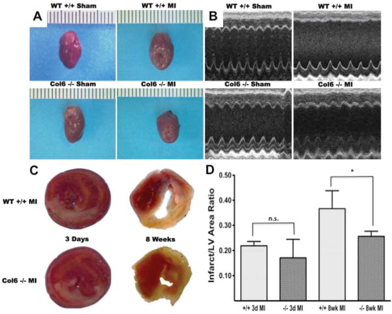
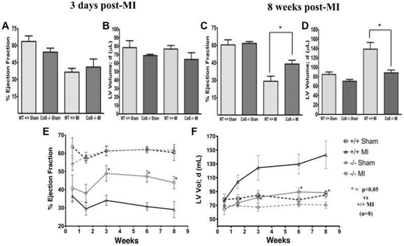
Methods and results: Wild-type and Col6a1(-/-) mice were subjected to MI, followed by serial echocardiographic and histological assessments. At 8 weeks after MI, infarct size was significantly reduced, ejection fraction was significantly preserved (43.9% ± 3.3% versus 29.1% ± 4.3% for wild-type), and left ventricular chamber dilation was attenuated in the Col6a1(-/-) MI group (25.8% ± 7.9% increase versus 62.6% ± 16.5% for wild-type). The improvement in cardiac remodeling was evident as early as 10 days after MI in the Col6a1(-/-) mice. Myocyte apoptosis within the infarcted zones was initially greater in the Col6a1(-/-) group 3 days after MI, but by day 14 this was significantly reduced. Collagen deposition also was reduced in the infarcted and remote areas of the Col6a1(-/-) hearts. The reductions in chronic myocyte apoptosis and fibrosis are critical events leading to improved long-term remodeling and functional outcomes.
Conclusions: These unexpected results demonstrate for the first time that deletion of type VI collagen in this knockout model plays a critical protective role after MI by limiting infarct size, chronic apoptosis, aberrant remodeling, and fibrosis, leading to preservation of cardiac function.
Figures

References
-
- Lindsey ML, Mann DL, Entman ML, Spinale FG. Extracellular matrix remodeling following myocardial injury. Ann Med. 2003;35:316–26. - PubMed
-
- Sun Y, Weber KT. Infarct scar: a dynamic tissue. Cardiovasc Res. 2000;46:250–6. - PubMed
-
- Naugle JE, Olson ER, Zhang X, Mase SE, Pilati CF, Maron MB, Folkesson HG, Horne WI, Doane KJ, Meszaros JG. Type VI collagen induces cardiac myofibroblast differentiation: implications for postinfarction remodeling. Am J Physiol Heart Circ Physiol. 2006;290:H323–30. - PubMed
Publication types
MeSH terms
Substances
Grants and funding
LinkOut - more resources
Full Text Sources
Other Literature Sources
Medical
Molecular Biology Databases
