. 2012 Feb 15;482(7386):529-33.
doi: 10.1038/nature10825.

Clonal selection drives genetic divergence of metastatic medulloblastoma

Affiliations

Clonal selection drives genetic divergence of metastatic medulloblastoma

Xiaochong Wu et al. Nature. .

Abstract

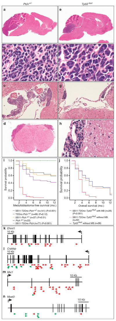
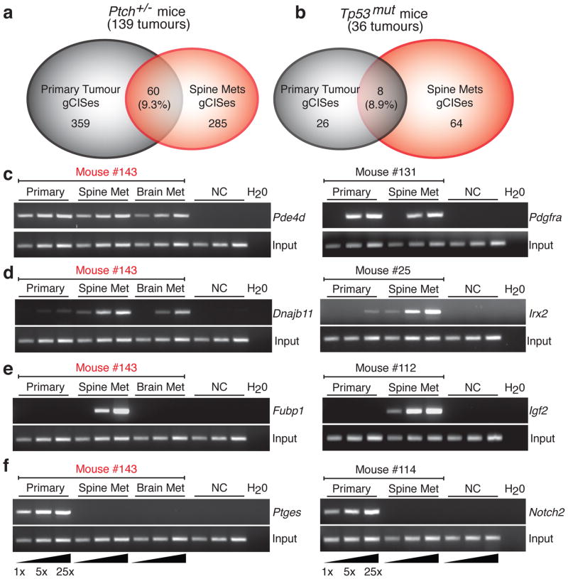
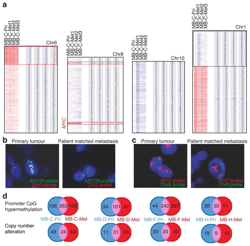
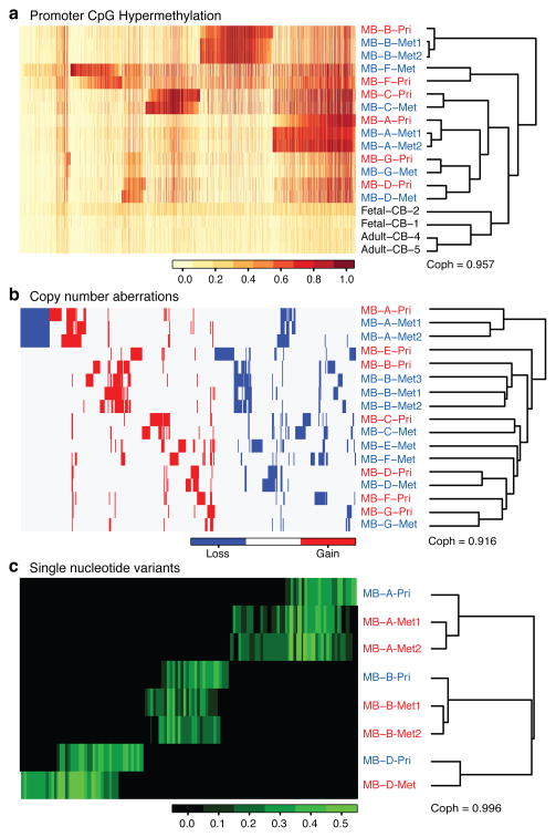
Medulloblastoma, the most common malignant paediatric brain tumour, arises in the cerebellum and disseminates through the cerebrospinal fluid in the leptomeningeal space to coat the brain and spinal cord. Dissemination, a marker of poor prognosis, is found in up to 40% of children at diagnosis and in most children at the time of recurrence. Affected children therefore are treated with radiation to the entire developing brain and spinal cord, followed by high-dose chemotherapy, with the ensuing deleterious effects on the developing nervous system. The mechanisms of dissemination through the cerebrospinal fluid are poorly studied, and medulloblastoma metastases have been assumed to be biologically similar to the primary tumour. Here we show that in both mouse and human medulloblastoma, the metastases from an individual are extremely similar to each other but are divergent from the matched primary tumour. Clonal genetic events in the metastases can be demonstrated in a restricted subclone of the primary tumour, suggesting that only rare cells within the primary tumour have the ability to metastasize. Failure to account for the bicompartmental nature of metastatic medulloblastoma could be a major barrier to the development of effective targeted therapies.


Conflict of interest statement

Author Information: The authors declare no competing financial interests.

Figures

Figure 1
Figure 1. Transposon mutagenesis models disseminated human medulloblastoma
Histology of transposon driven medulloblastoma on the Ptch +/− background (a to d) resembles human medulloblastoma with leptomeningeal metastases on the surface of the brain (c) and spinal cord (d). Histology of transposon driven medulloblastoma on the Tp53 mut background (e to h) show histological features of large cell/anaplastic medulloblastoma including nuclear pleomorphism and nuclear wrapping (f). Dissemination to the leptomeningeal spaces of the brain (g) and spinal cord (h). (i) Ptch +/− mice with sleeping beauty transposition develop more frequent medulloblastomas with a shorter latency than Ptch +/− mice without transposition (P values are from t-tests of survival comparing individual genotypes to Ptch +/− mice; n= # of mice per genotype). (j) Medulloblastoma was not seen in Tp53mut mice without transposition, but was seen in 42% of Tp53mut mice with transposition. (P values are from t-tests comparing survival between Tp53mut mice and SB11-T2Onc-Tp53mut and Tp53mut mice; n = # of mice). (k–n). Insertion maps of notable gCISes. Insertions in the direction of transcription denoted by green arrows, those against the direction of transcription by red arrows.
Figure 2
Figure 2. Transposon driven metastatic medulloblastoma genetically differs from the primary tumour
(a,b) Venn diagram showing the extent of overlap between gCISes in primary tumours and metastases on the Ptch +/− and Tp53mut backgrounds. (c–f) Insertion site, end point PCR to demonstrate relative clonality of insertions between samples. Three levels of input DNA were used for each sample as illustrated (1x, 5x, 25x). Insertions in the first column are from medulloblastoma #143 (c) Clonal events found in both primary tumour and matching metastases. (d) Insertions that are highly clonal in the metastases, but very sub-clonal in the matching primary tumour. (e) Insertions that are highly clonal in the metastases, but completely undetectable in the matching primary tumour. (f) Insertions that are highly clonal in the primary tumour, but completely undetectable in the matching metastases.
Figure 3
Figure 3. Human medulloblastoma metastases are biologically distinct from their matched primary tumour
(a) Copy number data from a primary medulloblastoma and three patient matched metastases, with chromosomal regions in red representing genetic gain (amplification), and blue denoting genetic loss (deletion). Examples of shared clonal events (red boxes), events limited to the metastases (blue boxes), and events limited to one but not all metastases (black box) are shown. (b) Interphase FISH shows amplification of MYCN in a primary tumour, but not the matched metastasis. (c) Interphase FISH for MYC demonstrates amplification in both the primary tumour and its matched metastases. (d) Venn diagrams depicting the degree of overlap and discordance in promoter CpG methylation events (above) and copy number alterations (below) in primary medulloblastomas and their matched metastasis.
Figure 4
Figure 4. Human medulloblastoma metastases are genetically distinct from the primary tumour
(a) Profiling the methylation status of 27,578 CpG dinucleotides sites in the human genome in a collection of human matched primary and metastatic medulloblastomas, top 2000 genes are shown. Unsupervised hierarchical clustering by CpG methylation patterns demonstrates that patient matched metastases are more similar to each other than to their matched primary tumour. (b) Unsupervised clustering of regions of copy number gain and loss demonstrates that patient matched metastases are more similar to each other, than they are to their matched primary tumour. (c) Unsupervised hierarchical clustering of SNV data from whole exome sequencing demonstrates that patient matched metastases are more similar to each other than to the patient matched primary tumour. SNVs that are found only in the primary compartment, or only in both tumours in the metastatic compartment are evident.

Comment in

References

    1. Gajjar A, et al. Risk-adapted craniospinal radiotherapy followed by high-dose chemotherapy and stem-cell rescue in children with newly diagnosed medulloblastoma (St Jude Medulloblastoma-96): long-term results from a prospective, multicentre trial. Lancet Oncol. 2006;7:813–820. doi: 10.1016/S1470-2045(06)70867-1. S1470-2045(06)70867-1 [pii] - DOI - PubMed
    1. Mabbott DJ, et al. Serial evaluation of academic and behavioral outcome after treatment with cranial radiation in childhood. J Clin Oncol. 2005;23:2256–2263. doi: 10.1200/JCO.2005.01.158. 23/10/2256 [pii] - DOI - PubMed
    1. MacDonald TJ, et al. Expression profiling of medulloblastoma:PDGFRA and the RAS/MAPK pathway as therapeutic targets for metastatic disease. Nat Genet. 2001;29:143–152. doi: 10.1038/ng731. ng731 [pii] - DOI - PubMed
    1. Ramaswamy S, Ross KN, Lander ES, Golub TR. A molecular signature of metastasis in primary solid tumors. Nat Genet. 2003;33:49–54. doi: 10.1038/ng1060. ng1060 [pii] - DOI - PubMed
    1. Goodrich LV, Milenkovic L, Higgins KM, Scott MP. Altered neural cell fates and medulloblastoma in mouse patched mutants. Science. 1997;277:1109–1113. - PubMed

Publication types

Substances

Associated data