. 2012 Feb 19;483(7387):92-5.
doi: 10.1038/nature10844.

Repetitive motor learning induces coordinated formation of clustered dendritic spines in vivo

Affiliations

Repetitive motor learning induces coordinated formation of clustered dendritic spines in vivo

Min Fu et al. Nature. .

Abstract

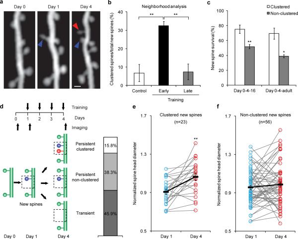
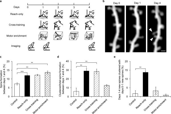
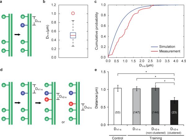
Many lines of evidence suggest that memory in the mammalian brain is stored with distinct spatiotemporal patterns. Despite recent progresses in identifying neuronal populations involved in memory coding, the synapse-level mechanism is still poorly understood. Computational models and electrophysiological data have shown that functional clustering of synapses along dendritic branches leads to nonlinear summation of synaptic inputs and greatly expands the computing power of a neural network. However, whether neighbouring synapses are involved in encoding similar memory and how task-specific cortical networks develop during learning remain elusive. Using transcranial two-photon microscopy, we followed apical dendrites of layer 5 pyramidal neurons in the motor cortex while mice practised novel forelimb skills. Here we show that a third of new dendritic spines (postsynaptic structures of most excitatory synapses) formed during the acquisition phase of learning emerge in clusters, and that most such clusters are neighbouring spine pairs. These clustered new spines are more likely to persist throughout prolonged learning sessions, and even long after training stops, than non-clustered counterparts. Moreover, formation of new spine clusters requires repetition of the same motor task, and the emergence of succedent new spine(s) accompanies the strengthening of the first new spine in the cluster. We also show that under control conditions new spines appear to avoid existing stable spines, rather than being uniformly added along dendrites. However, succedent new spines in clusters overcome such a spatial constraint and form in close vicinity to neighbouring stable spines. Our findings suggest that clustering of new synapses along dendrites is induced by repetitive activation of the cortical circuitry during learning, providing a structural basis for spatial coding of motor memory in the mammalian brain.


Figures

Figure 1
Figure 1. Acquisition of a novel motor skill induces formation of spine clusters
a, Repeated imaging of the same dendritic branch during motor learning reveals that a second new spine that formed between days 1 and 4 (red arrowhead) is located next to a stabilized new spine that had formed on day 1 (blue arrowhead). Scale bar, 1 μm. b, A higher percentage of new spines formed in clusters over 4 days during early training (n=18 mice), compared to control (n=7) and late training (n=4). c, Clustered new spines observed on training day 4 have a higher survival rate than non-clustered counterparts by the end of the 16-day training (n=6), as well as 4 months after training stops (n=4). d, New spines formed on training day 1 are classified according to their fate and neighboring spine formation. e, Spine head sizes of persistent clustered new spines increase between training days 1 and 4. f, Spine head sizes of persistent non-clustered new spines show no change between training days 1 and 4. Spine head size is quantified by the normalized spine head diameter, defined as the ratio of the spine head diameter to the adjacent dendritic shaft diameter. *P<0.05, **P<0.01. Error bars, s.e.m.
Figure 2
Figure 2. Clustered new spines form over multiple training sessions of the same, but not different, motor tasks
a, Timelines of reach-only, cross-training and motor enrichment experiments. b, Repeated imaging of the same dendritic branch revealed that two neighboring new spines (arrowhead) formed between days 1 and 4 during cross-training. Scale bar, 1 μm. c, Higher percentages of new spines formed between days 1 and 4 in reach-only, cross-training and motor enrichment, compared to controls. d, Higher percentages of new spines formed in clusters between days 1 and 4 in reach-only and cross-training, compared to controls. e, A higher percentage of new spines that formed between days 1 and 4 clustered with new spines that had formed between days 0 and 1 in the reach-only condition, compared to controls. Number of mice examined: 6 control, 9 reach-only, 5 cross-training and 6 motor enrichment. **P<0.01, ***P<0.001. Error bars, s.e.m.
Figure 3
Figure 3. The spatial distribution of new spines along dendrites
a, A schematic illustrating the measurement of Dn-s. b, The median of measured Dn-ss (the red circle) is significantly larger than that of simulated Dn-ss (box plot of results from 1000 simulations, with whiskers representing the minimum and the maximum) in control mice. The simulation is based on the null hypothesis that new spines are added independently and uniformly along a linear dendrite. c, Cumulative probability distribution of measured Dn-s is shifted towards longer distances than the simulated Dn-s in control mice. d, A schematic illustrating the measurement of Dn2-s, clustered. The nearest spine to a clustered n2 could be either a persistent first new spine (n1) or a stable spine existing since day 0, depending on relative n2 location. e, Dn1-s in control mice is comparable to that of trained mice. In trained mice, Dn2-s, clustered is significantly smaller than Dn1-s, while Dn2-s, non-clustered is comparable to Dn1-s. The number of spines analyzed in each condition is indicated on each column. *P<0.05. Error bars, s.e.m.

References

    1. Silva AJ, Zhou Y, Rogerson T, Shobe J, Balaji J. Molecular and cellular approaches to memory allocation in neural circuits. Science. 2009;326:391–395. - PMC - PubMed
    1. Aimone JB, Wiles J, Gage FH. Potential role for adult neurogenesis in the encoding of time in new memories. Nature Neurosci. 2006;9:723–727. - PubMed
    1. Han JH, et al. Selective erasure of a fear memory. Science. 2009;323:1492–1496. - PubMed
    1. Komiyama T, et al. Learning-related fine-scale specificity imaged in motor cortex circuits of behaving mice. Nature. 2010;464:1182–1186. - PubMed
    1. Kee N, Teixeira CM, Wang AH, Frankland PW. Preferential incorporation of adult-generated granule cells into spatial memory networks in the dentate gyrus. Nature Neurosci. 2007;10:355–362. - PubMed

Publication types