. 2012 Feb 19;14(3):318-28.
doi: 10.1038/ncb2426.

A genome-wide homologous recombination screen identifies the RNA-binding protein RBMX as a component of the DNA-damage response

Affiliations

A genome-wide homologous recombination screen identifies the RNA-binding protein RBMX as a component of the DNA-damage response

Britt Adamson et al. Nat Cell Biol. .

Abstract

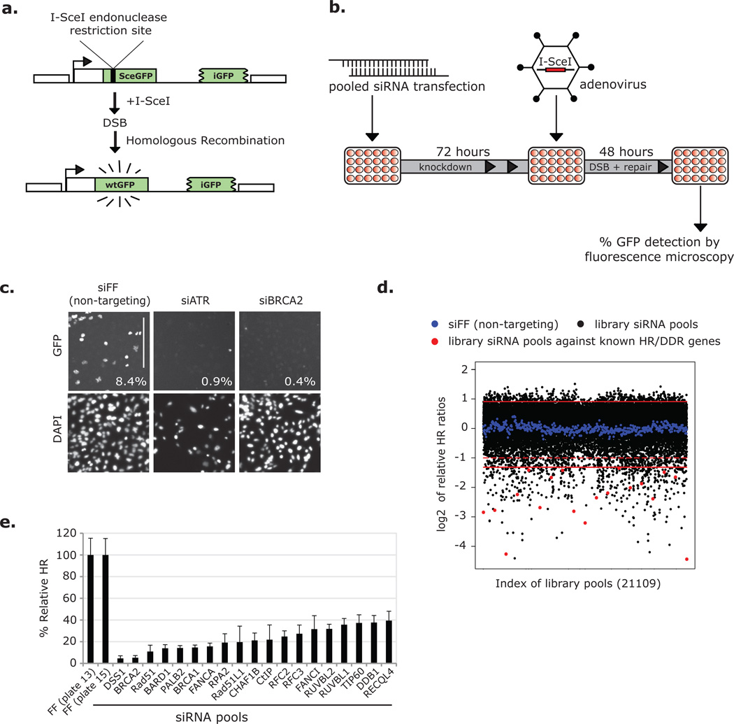
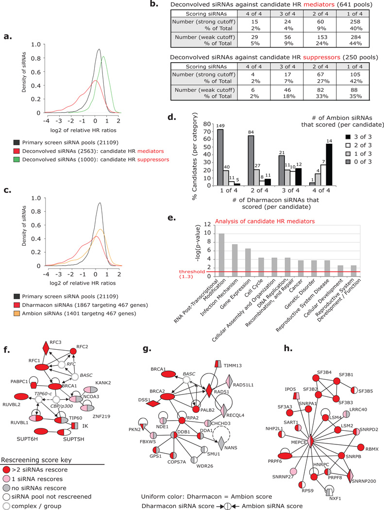
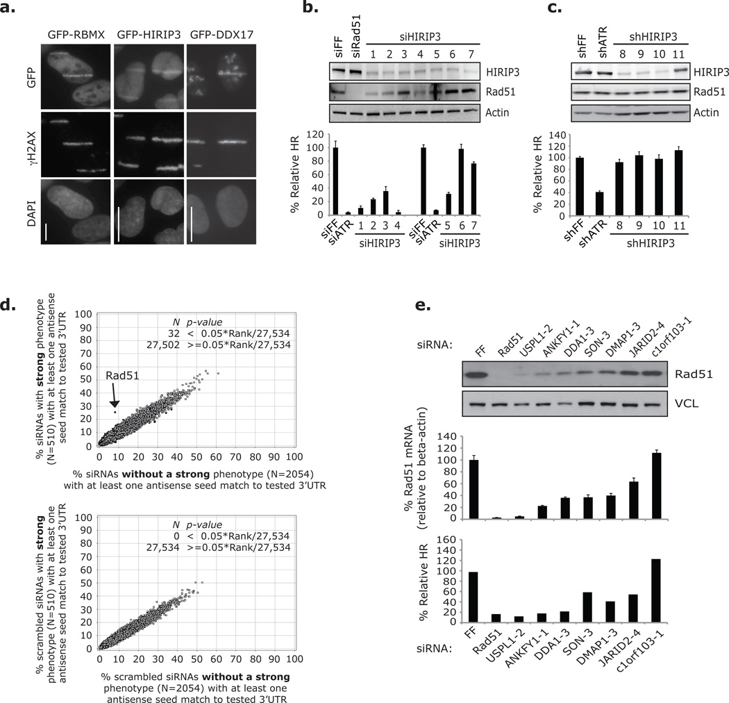
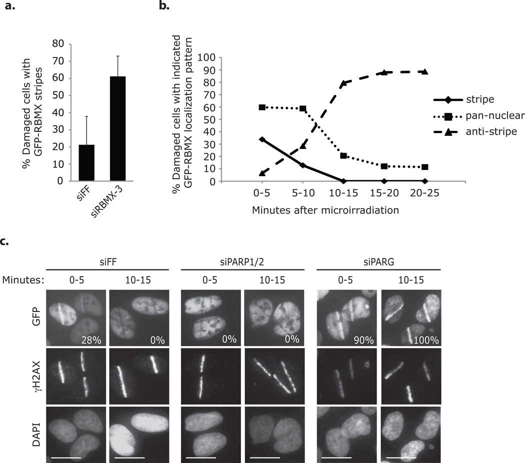
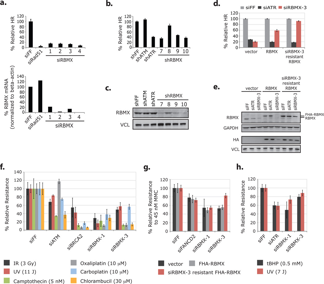
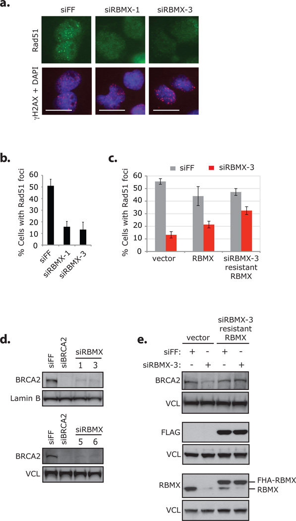
Repair of DNA double-strand breaks is critical to genomic stability and the prevention of developmental disorders and cancer. A central pathway for this repair is homologous recombination (HR). Most knowledge of HR is derived from work in prokaryotic and eukaryotic model organisms. We carried out a genome-wide siRNA-based screen in human cells. Among positive regulators of HR we identified networks of DNA-damage-response and pre-mRNA-processing proteins, and among negative regulators we identified a phosphatase network. Three candidate proteins localized to DNA lesions, including RBMX, a heterogeneous nuclear ribonucleoprotein that has a role in alternative splicing. RBMX accumulated at DNA lesions through multiple domains in a poly(ADP-ribose) polymerase 1-dependent manner and promoted HR by facilitating proper BRCA2 expression. Our screen also revealed that off-target depletion of RAD51 is a common source of RNAi false positives, raising a cautionary note for siRNA screens and RNAi-based studies of HR.


Figures

Figure 1
Figure 1
A genome-wide siRNA-based screen for homologous recombination (HR) genes. (a) Schematic of DR-GFP construct. (b) Schematic of the high-throughput (HTP) HR screen. Arrayed pools of siRNAs were reverse transfected into DR-U2OS cells in 384 well plates. Cells were infected with the I-SceI expressing adenovirus AdNGUS24i at an MOI of ~10 after 72 hours and 48 hours later were fixed and imaged. (c) DR-U2OS cells were transfected with positive (siATR and siBRCA2) and negative (siFF) control siRNAs and assayed for HR in high throughput. Images were taken on the automated screening platform and are presented in representative portions. % GFP+ cells –as calculated from full images– are included. Scale bar represents 300 µm. (d) Relative HR ratios (presented as log2 values) from 22109 library siRNA pools and 476 negative controls (siFF). Solid red lines indicate 2 s.d. from the screen-wide mean of relative HR ratios (presented as log2 values). These were used as cutoff values to determine pools scoring with increased or decreased HR. The region between the dotted red line and the lower solid red line indicates a scoring range from which most additional candidate HR mediators were selected. (e) Known regulators of HR or the DDR that scored under the 2 s.d.-based cutoff.
Figure 2
Figure 2
Rescreen and validation of candidate HR genes. (a) The distributions of relative HR ratios (as log2 values) for three sets of screened siRNAs: primary screen pools (black), deconvolved Dharmacon siRNAs against candidate HR mediators (red), and deconvolved Dharmacon siRNAs against candidate HR suppressors (green). (b) The number and percentage of Dharmacon siRNA pools that rescored with 1, 2, 3 or 4 siRNAs after deconvolution. Results analyzed using strong (2 s.d. from the screen-wide mean-based: 40% and 188% relative HR) or weak (1.5 s.d.-based: 59% and 169%) cutoff values. (c) The distributions of relative HR ratios (as log2 values) for primary screen pools (black: as presented in a), siRNAs from select Dharmacon pools against candidate mediators (red), and rescreened Ambion siRNAs against candidate mediators (orange). Dharmacon and Ambion siRNAs target the same 467 candidate genes. (d) Percentage of candidate HR mediator genes that scored with 0, 1, 2, or 3 Ambion siRNAs in categories of those that scored with 1, 2, 3, or 4 Dharmacon siRNAs. Weak cutoff was used for scoring. Number of candidates indicated. (e) Functional gene categories enriched among candidate HR mediators identified using Ingenuity Pathway Analysis (IPA). (f–h) Interaction networks generated from candidates that scored as HR mediators in the primary screen identified by IPA: (f) DDR network including components of the TIP60 complex and the RFC DNA clamp loader, (g) DDR / HR network including canonical HR proteins and Cul4A ubiquitin ligase associated proteins, (h) pre-mRNA processing network. Color key (representative of rescreening data): red indicates a candidate that rescored with >2 siRNAs (out of 4 Dharmacon and 3 Ambion), pink indicates that 1 siRNA rescored, gray indicates that 0 siRNAs rescored, and white indicates a candidate that was not rescreened. Line key: solid lines indicate direct interactions, dashed lines indicate indirect actions, arrows indicate the direction of interactions, and lines without arrowheads indicate binding. Shape key: ovals indicate transcription regulators, hexagons indicate translational regulators, diamonds (vertical) indicate enzymes, diamonds (horizontal) indicate peptidases, trapezoids indicate transporters, inverted triangles indicate kinases, squares indicate cytokines, circles indicate other, double circles indicate groups of proteins or complexes.
Figure 3
Figure 3
Off-target Rad51-depletion was a major source of false positives among Dharmacon siRNAs identified by the primary screen. (a) Cells expressing GFP fusions of RBMX, HIRIP3 or DDX17 were microirradiated and prepared for imaging after 0–5 (RBMX and DDX17) and 15–20 minutes (HIRIP3) with an antibody against γH2AX. GFP was observed directly. Nuclei were stained with DAPI. Scale bars indicate 10 µm. Images were prepared from three separate experiments and are not intended for comparison. (b) HR assay results from DR-U2OS and HIRIP3 / Rad51 western blot analysis from U2OS cells transfected with siRNAs against HIRIP3 in three separate experiments: one to acquire western blot data and two for HR analysis. Experimental data is grouped with corresponding controls. Error bars represent ± s.d. across three replicates. (c) HR assay results and corresponding HIRIP3 / Rad51 western blot analysis from DR-U2OS cells transduced with shRNAs against HIRIP3. Error bars represent ± s.d. across three replicates. (d) GESS analysis of Dharmacon siRNAs against candidate mediator genes. Scatter plots represent the percentage of siRNAs in 2 groups that have at least one 7-nucleotide antisense seed sequence match to 27,534 human 3’UTRs. Upper plot: compares siRNAs that individually rescored with a strong phenotype (y-axis) to those that did not (x-axis). Lower plot: compares the same 2 groups of siRNAs after scrambling the seed sequences and serves as a control. 3’UTRs that significantly enriched for seed matches in either siRNA group are black. Arrow illustrates the 3’UTR of Rad51. (e) HR assay results and Rad51 mRNA / protein levels from cells transfected with seven siRNAs predicted to off-target Rad51 by a 7-nucleotide antisense seed region match to the Rad51 3’UTR. Western blot and RT-qPCR results are from the same experiment in U2OS cells. Primers used against Rad51 mRNA recognize four transcript variants. Error bars in RT-qPCR analysis represent ± s.e.m. across three replicates. HR data is from the HTP Dharmacon rescreening analysis in DR-U2OS cells and represent the average of two replicates. Full scans of blots in b–c, e are shown in Supplementary Information, Fig. S13.
Figure 4
Figure 4
RBMX accumulates transiently at sites of DNA damage in a PARP-dependent manner. (a) U2OS cells expressing siRBMX-3 resistant GFP-RBMX and transfected with the indicated siRNAs were microirradiated (one at a time for 5 minutes) and then immediately processed for immunofluorescence with an antibody against γH2AX. Cells were evaluated for GFP-RBMX accumulation at γH2AX stained laser tracks (approx. 60–190 cells / condition). Error bars represent ± s.d. across three replicates. (b) U2OS cells expressing GFP-RBMX were microirradiated (one at a time for 5 minutes) and processed for immunofluorescence with an antibody against γH2AX at the indicated times. Cells were evaluated for GFP-RBMX accumulation at γH2AX stained laser tracks (approx. 130–190 cells / condition). Data represent the mean of two replicates. (c) U2OS cells expressing GFP-RBMX and transfected with the indicated siRNAs were microirradiated (one at a time for 5 minutes) and processed for imaging with an antibody against γH2AX at the indicated times. GFP was observed directly. Nuclei were stained with DAPI. The percentages of cells with GFP-RBMX accumulation at γH2AX stained laser tracks are indicated (approx. 110–160 cells / condition, n=1). siPARP1/2 indicates two pools of 4 siRNAs targeting PARP1 and PARP2. siPARG indicates a pool of 4 siRNAs targeting PARG. Scale bars indicate 10 µm.
Figure 5
Figure 5
RBMX promotes homologous recombination and resistance to DNA damaging agents. (a) HR assay results from DR-U2OS cells transfected with RBMX-targeting siRNAs and corresponding levels of RBMX mRNA as measured by RT-qPCR (normalized to beta-actin mRNA). Error bars in HR analysis represent ± s.d. across three replicates and data in RT-qPCR analysis represent the mean of two replicates. (b) HR assay results from DR-U2OS cells transduced with RBMX-targeting shRNAs. Error bars represent ± s.d. across three replicates. (c) Western blot analysis of RBMX levels from cells analyzed in b. (d) DR-U2OS cells transduced with control vector, FHA-RBMX and siRBMX-3 resistant FHA-RBMX cDNAs and then transfected with indicated siRNAs were assayed for HR efficiency. Data from cells with the same cDNA were normalized to the siFF condition. Error bars represent ± s.d. across three replicates. (e) Whole-cell extracts from cells in d were immunoblotted with the indicated antibodies. Two western blots of the same extracts are presented and panels are grouped accordingly. (f) Multicolor competition assay for resistance to DNA damaging agents. Briefly, U2OS cells expressing GFP and transfected with the indicated siRNAs were mixed in a 1:1 ratio with dsRed U2OS cells transfected with siFF. Cell mixtures were treated with the indicated DNA damaging agents, and after 8 days, the relative survival of GFP to dsRed cells was determined by FACS analysis. GFP / dsRed ratios were normalized to those from undamaged pools to control for the relative growth effects of the siRNAs. Error bars represent ± s.d. across three replicates. (g) DR-U2OS cells transduced with the indicated cDNAs and then transfected with the indicated siRNAs were treated with 45 nM MMC, and after 7 days relative resistance was determined by the CellTiter-Glo Luminescent Cell Viability Assay. Error bars represent ± s.d. across three replicates. (h) U2OS cells transfected with the indicated siRNAs were treated with tBHP (for 1 hour) or UV, and after 5 days relative resistance was determined by the CellTiter-Glo Luminescent Cell Viability Assay. Error bars represent ± s.d. across three replicates. Full scans of blots in c and e are shown in Supplementary Information, Fig. S13.
Figure 6
Figure 6
RBMX promotes formation of IR-induced Rad51 foci by facilitating proper expression of BRCA2. (a) DR-U2OS cells were transfected with the indicated siRNAs, treated with 10 Gy IR, and processed for imaging with antibodies against Rad51 and γH2AX after 4 hours. Nuclei were stained with DAPI. Scale bars indicate 20 µm. Data from the same experiment is presented in Supplementary Information, Fig. S7d–g, S8c. Individual adjustment of color channels in γH2AX + DAPI was required to illustrate foci; identical adjustment parameters were used. (b) DR-U2OS cells transfected with the indicated siRNAs were damaged and processed for immunofluorescence in same manner as described in a. Cells with Rad51 foci were counted by eye; because transfection with siRNAs against RBMX causes some changes to nuclear morphology, only normal shaped nuclei were counted (approx. 100–130 cells / condition). Error bars represent ± s.d. across four replicates. Data from the same siRNA transfected cells are presented in Supplementary Information, Fig S9b–c. (c) DR-U2OS cells transduced with control vector, FHA-RBMX or siRBMX-3 resistant FHA-RBMX cDNAs were transfected with indicated siRNAs and treated with 10 Gy IR. After 8 hours cells were processed for immunofluorescence with antibodies against Rad51 and γH2AX and counted (200 cells / condition). Error bars represent ± s.d. across four replicates. Representative images of cells are presented in Supplementary Information, Fig. S8d. (d) Whole-cell extracts from DR-U2OS cells transfected with the indicated siRNAs were immunoblotted with antibodies against BRCA2 or Lamin B / vinculin (VCL). Extracts analyzed in the upper blot were evaluated for protein levels of additional HR and DDR proteins (Supplementary Information, Fig. S12a); and additional data from this blot are presented in Supplementary Information, Fig. S12b (the Lamin B panel is reproduced as a control in that figure). Extracts analyzed in the lower blot were also used in Supplementary Information, Fig. S7b / S12d. (e) Whole-cell extracts from undamaged DR-U2OS cells evaluated in c were immunoblotted with the indicated antibodies. Three western blots of the same extracts are presented and panels are grouped accordingly. Full scans of blots in d–e are shown in Supplementary Information, Fig. S13.

References

    1. Ciccia A, Elledge SJ. The DNA damage response: making it safe to play with knives. Mol Cell. 2010;40:179–204. - PMC - PubMed
    1. Sartori AA, et al. Human CtIP promotes DNA end resection. Nature. 2007;450:509–514. - PMC - PubMed
    1. Bolderson E, et al. Phosphorylation of Exo1 modulates homologous recombination repair of DNA double-strand breaks. Nucleic Acids Res. 2010;38:1821–1831. - PMC - PubMed
    1. West SC. Molecular views of recombination proteins and their control. Nat Rev Mol Cell Biol. 2003;4:435–445. - PubMed
    1. San Filippo J, Sung P, Klein H. Mechanism of eukaryotic homologous recombination. Annu Rev Biochem. 2008;77:229–257. - PubMed

Publication types

MeSH terms