. 2012 Jun 1;125(Pt 11):2638-54.
doi: 10.1242/jcs.096875. Epub 2012 Feb 17.

Mammary collective cell migration involves transient loss of epithelial features and individual cell migration within the epithelium

Affiliations

Mammary collective cell migration involves transient loss of epithelial features and individual cell migration within the epithelium

Andrew J Ewald et al. J Cell Sci. .

Abstract

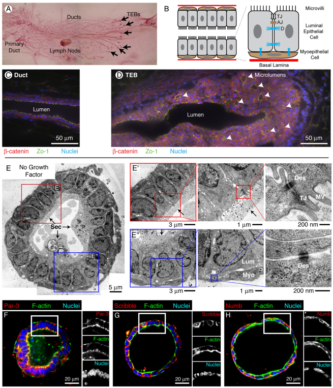
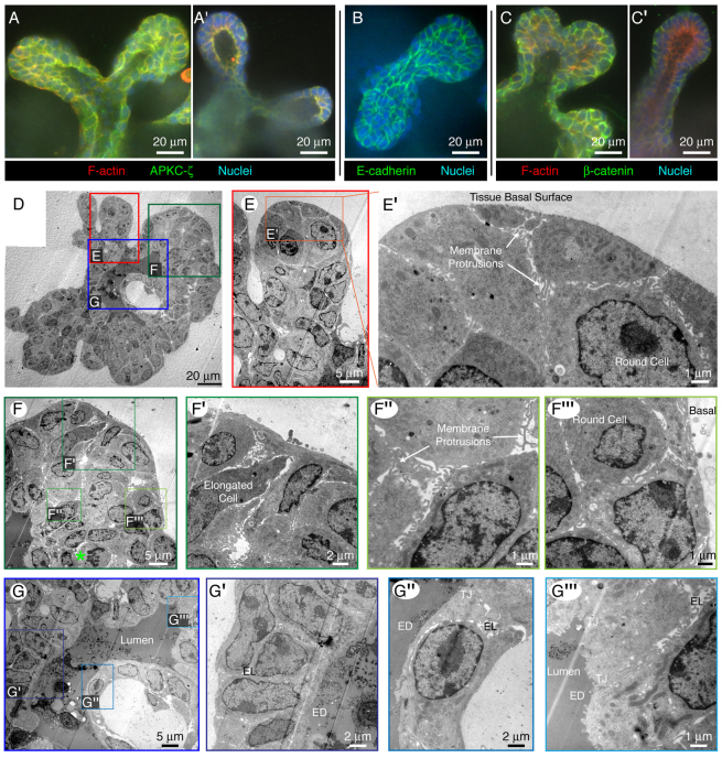
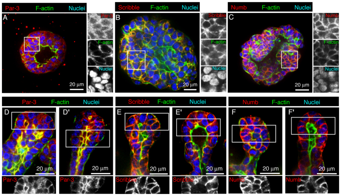
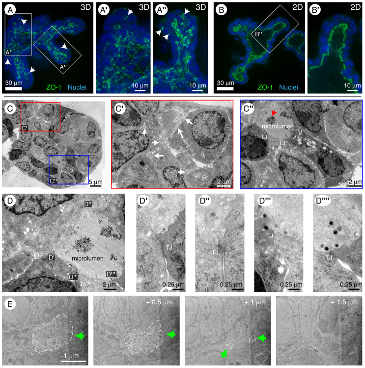
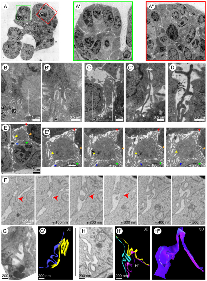
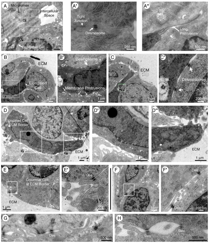
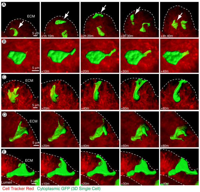
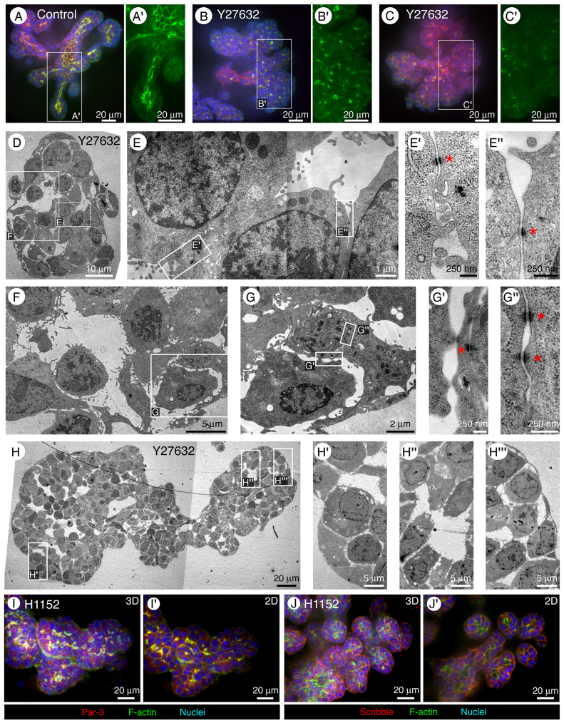
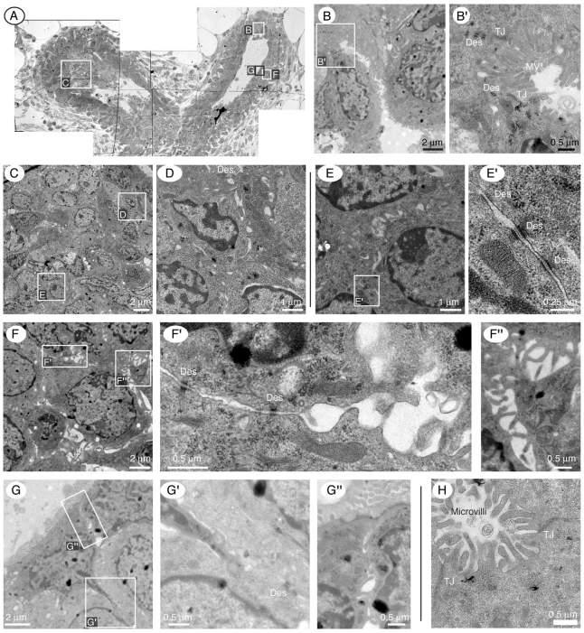
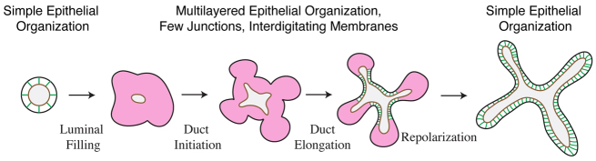
Normal mammary morphogenesis involves transitions between simple and multilayered epithelial organizations. We used electron microscopy and molecular markers to determine whether intercellular junctions and apico-basal polarity were maintained in the multilayered epithelium. We found that multilayered elongating ducts had polarized apical and basal tissue surfaces both in three-dimensional culture and in vivo. However, individual cells were only polarized on surfaces in contact with the lumen or extracellular matrix. The basolateral marker scribble and the apical marker atypical protein kinase C zeta localized to all interior cell membranes, whereas PAR3 displayed a cytoplasmic localization, suggesting that the apico-basal polarity was incomplete. Despite membrane localization of E-cadherin and β-catenin, we did not observe a defined zonula adherens connecting interior cells. Instead, interior cells were connected through desmosomes and exhibited complex interdigitating membrane protrusions. Single-cell labeling revealed that individual cells were both protrusive and migratory within the epithelial multilayer. Inhibition of Rho kinase (ROCK) further reduced intercellular adhesion on apical and lateral surfaces but did not disrupt basal tissue organization. Following morphogenesis, segregated membrane domains were re-established and junctional complexes re-formed. We observed similar epithelial organization during mammary morphogenesis in organotypic culture and in vivo. We conclude that mammary epithelial morphogenesis involves a reversible, spatially limited, reduction in polarity and intercellular junctions and active individualistic cell migration. Our data suggest that reductions in polarity and adhesion during breast cancer progression might reflect partial recapitulation of a normal developmental program.


Figures

Fig. 1.
Fig. 1.
Normal mammary morphogenesis is accomplished by a stratified epithelium. (A) Carmine-Red-stained 10-week-old mouse mammary gland. Mammary ducts are elongated during puberty by specialized structures at the end of the duct, terminal end buds (TEBs). (B) Resting mammary ducts have a bilayered organization, with luminal epithelial cells, connected by extensive intercellular junctions, and basally located myoepithelial cells. (C) Normal ducts in vivo have a simple epithelial organization when not actively growing. Zona occludens 1 (ZO-1) is localized to the apico-lateral surface of the luminal epithelial cells and β-catenin to the basolateral surfaces. (D) Mammary ducts are elongated during puberty by TEBs, stratified epithelial structures with many luminal cell layers. β-catenin localizes to all basolateral surfaces and is only excluded from lumen-facing surfaces. ZO-1 localizes to the lumen lining surfaces of both the main lumen and isolated micro-lumens. (EE″) Primary mammary ducts can be isolated and grown in 3D Matrigel gels. Without the addition of growth factor all ducts form simple cysts. These cysts are bilayered, with a single luminal (Lum) cell layer and a single myoepithelial (Myo) layer. The lumen has electron dense secretory material (arrows), microvilli (MV), and tight junctions (TJ). Both luminal and myoepithelial cells are connected by desmosomes (Des). (FH) Polarized cysts in 3D culture localize PAR3 to apical surfaces (F) and both scribble (G) and numb (H) to basolateral surfaces. All TEM images are from high-pressure frozen, freeze-substituted samples that were pre-fixed with 4% glutaraldehyde.
Fig. 2.
Fig. 2.
During morphogenesis in 3D culture the mammary epithelium is transiently stratified. (AC′) Within the multilayered epithelium of elongating buds in 3D culture, most interior cell surfaces are not in contact with the lumen or the ECM. These interior surfaces stained positive for aPKC-ζ (A), E-cadherin (B) and β-catenin (C). Following morphogenesis, the mammary epithelium regained a simple organization. In regions of simple organization, aPKC-ζ localized to apical domains (A′) and β-catenin localized to basolateral domains (C′). (DE′) TEM was used to define the ultrastructural polarity of the tissue. The basal tissue surface was smooth and lacks ECM-directed protrusions. (FG‴) Both elongated cells (F′) and round unpolarized cells (F‴) are present in the interior of the multilayer. Away from the basal tissue surface, the epithelial cells exhibit dense, inter-digitating membrane extensions (F″). We also observed division of round cells distant from either the ECM or lumen facing surfaces (F, green asterisk). (GG‴) Within the same branching structure, there are regions with simple epithelial organization with an electron-dense lumen and tight junctions. All TEM images are from high-pressure frozen, freeze-substituted samples that were pre-fixed with 4% glutaraldehyde. ED, electron dense; EL, electron lucid; TJ, tight junction.
Fig. 3.
Fig. 3.
Morphogenesis in 3D culture is associated with a loss of molecular polarity. (AC) Interior cells are first evident in ‘complex cyst’ structures. Within these structures interior cells display cytoplasmic localization of PAR3 (A), whereas scribble and numb localize to all interior cell surfaces (B,C). (DF′) In the multilayered region of elongating mammary end buds PAR3 is cytoplasmically localized (D), whereas scribble and numb localize to all interior surfaces. After morphogenesis is complete the epithelium regains a simple epithelial organization and PAR3 is associated with the apical membrane (D′), whereas scribble and numb are localized to basolateral cell surfaces (E′,F′).
Fig. 4.
Fig. 4.
The multilayered region contains microlumens with tight junctions. (AA″) Light microscopy of the multilayered region. ZO-1 (green) localizes both to the lining of the main lumen and to isolated microlumens located between luminal epithelial cells (arrowheads in 3D volume reconstruction). (B,B′) Following morphogenesis a simple epithelial organization is restored and a single clear lumen is observed. (CD‴) Using TEM there are two different intercellular spaces: electron-lucid spaces with extensive irregular membrane protrusions but without tight junctions (C′, white arrows) and electron-dense intercellular spaces with tight junctions (TJ), microvilli and secretory material (red arrow in C″) (C″–D‴). (E) Serial block face scanning electron microscopy enabled 3D reconstruction of the microlumens and confirmed that they are several microns thick. All TEM images are from high-pressure frozen, freeze-substituted samples that were pre-fixed with 4% glutaraldehyde. The image series used to generate E has been uploaded to ‘The Cell: An Image Library’.
Fig. 5.
Fig. 5.
Cell shape and lateral membrane organization are highly heterogeneous in the multilayered region. (AA″) Nearby regions in the same branching structure can have high (A′) or low (A″) epithelial organization. (BD) Lateral membrane protrusions within the multilayer were frequently interdigitated (B–B′), could be several microns in length (C–C′) and were frequently branched (D). (E–F) Analysis of serial sections by TEM (E,E′) and serial block face scanning electron microscopy (F) revealed that the lateral membrane protrusions extend through multiple sections (colored arrowheads) and can morph between thin and broad and between linear and branched. (GH″) Three-dimensional reconstructions of cell contact regions along interior lateral surfaces using serial block face SEM revealed densely interdigitating 3D membrane extensions. All TEM images are from high-pressure frozen, freeze-substituted samples that were pre-fixed with 4% glutaraldehyde. The image series used to generate F has been uploaded to ‘The Cell: An Image Library’.
Fig. 6.
Fig. 6.
Elongated cells are observed within the epithelial multilayer. (AA″) Electron-dense elongated cells are seen within the multilayered region. These cells frequently have long protrusions extending in a single direction between adjacent cells (A″). (B–C′) Elongated cells were observed deep within the multilayer (A) and also within microns of the ECM (B). The only intercellular junctions observed on these cells were small desmosomes (CC′). (D–D″) At the ECM border some cells had an appearance that was intermediate between columnar epithelial and elongated morphologies. They did not extend protrusions into the ECM. (EF′) Cells in extensive contact with the ECM had smooth basal surfaces and lateral desmosomes, but frequently displayed little morphologic polarity on their lateral surfaces. (GH) Interior cells have lateral surfaces with intermixed membrane protrusions and small desmosomes. All TEM images are from high-pressure frozen, freeze-substituted samples that were pre-fixed with 4% glutaraldehyde.
Fig. 7.
Fig. 7.
Interior cells are frequently migratory and protrusive within the multilayer. All cells were labeled with Cell Tracker Red and a subset of cells were labeled with an adenovirally delivered cytoplasmic GFP. Images depict 3D reconstructions of the cytoplasm in individual GFP-positive cells. (A) Interior cells were frequently highly migratory and could move in directions opposite to the direction of ductal elongation. (B) Interior cells extended and retracted cytoplasmic protrusions within the multilayer, but not into the lumen or ECM. (C) Individual cells migrated from the interior to basal positions in contact with the ECM, a process termed radial intercalation. (D,E) Cells in contact with the basal tissue surface (D) or both apical and basal tissue surfaces (D,E) extended cytoplasmic protrusions from all lateral surfaces. The movies used to generate A–E are presented in supplementary material Movies 1–5, respectively.
Fig. 8.
Fig. 8.
Treatment with Y-27632 results in disorganization and reduced cell–cell contact on lateral and apical surfaces. (A–C′) Normal organoids have a stereotyped branching pattern, with a large ZO-1-lined lumen (A,A′). Treatment with the ROCK inhibitor Y-27632 results in rapid loss of the lumen, luminal epithelial disorganization and localization of ZO-1 exclusively to small foci (B,B′). Large regions of Y-27632-treated epithelium were free of ZO-1 immunoreactivity (red, phalloidin; green, ZO-1; blue, nuclei). (DH‴) Ultrastructural examination revealed a loss of electron-dense luminal spaces and a large decrease in cell–cell contact along lateral cell surfaces. The basal tissue surface was still smooth and well organized (H–H‴). The only junctions we observed connecting cells in Y-27632-treated samples were small desmosomes (red asterisks, E′,E″,G′,G″). All TEM images are from high-pressure frozen, freeze-substituted samples that were pre-fixed with 4% glutaraldehyde. (I,I′) Organoids treated with the ROCK inhibitor H1152 had microlumen-localized PAR3 (I, 3D reconstruction; I′, 2D optical section). (J,J′) Organoids treated with H1152 had lateral-surface-localized scribble but no scribble on the microlumen surface (J, 3D reconstruction; J′, 2D optical section).
Fig. 9.
Fig. 9.
TEBs in vivo display reduced apico-basal polarity and extensive intercellular membrane protrusions. (A) Mammary epithelial ducts are elongated by TEBs, shown here in a light micrograph, with boxes indicating regions that were subsequently imaged by TEM. (B,B′) TEBs contain a fluid-filled lumen with microvilli and tight junctions. (CF″) Cells within the multilayered region appeared morphologically unpolarized and displayed extensive intercellular protrusions. These protrusions were observed to interdigitate (D) and branch (F″). (GG″) Cells at the apical or basal most tissue surface were polarized at the tissue boundary but could be unpolarized and irregularly shaped a few microns away. (H) Microlumens within the body cell region had both microvilli and tight junctions. All TEM images are from high pressure frozen, freeze-substituted samples that were pre-fixed with 4% glutaraldehyde. Des, desmosomes; TJ, tight junctions.
Fig. 10.
Fig. 10.
Normal mammary epithelial morphogenesis is accomplished via a transiently stratified epithelium. The transient multilayered epithelium associated with mammary morphogenesis is polarized as a tissue but displays reduced apico-basal polarity and few intercellular junctions at points of cell–cell contact in the interior.

References

    1. Berx G., Cleton-Jansen A. M., Strumane K., de Leeuw W. J., Nollet F., van Roy F., Cornelisse C. (1996). E-cadherin is inactivated in a majority of invasive human lobular breast cancers by truncation mutations throughout its extracellular domain. Oncogene 13, 1919-1925 - PubMed
    1. Bilder D., Li M., Perrimon N. (2000). Cooperative regulation of cell polarity and growth by Drosophila tumor suppressors. Science 289, 113-116 - PubMed
    1. Bogenrieder T., Herlyn M. (2003). Axis of evil: molecular mechanisms of cancer metastasis. Oncogene 22, 6524-6536 - PubMed
    1. Cardona A., Saalfeld S., Preibisch S., Schmid B., Cheng A., Pulokas J., Tomancak P., Hartenstein V. (2010). An integrated micro- and macro-architectural analysis of the Drosophila brain by computer-assisted serial section electron microscopy. PLoS Biol. 8, e1000502 - PMC - PubMed
    1. Chepko G., Smith G. H. (1997). Three division-competent, structurally-distinct cell populations contribute to murine mammary epithelial renewal. Tissue Cell 29, 239-253 - PubMed

Publication types

MeSH terms