. 2012 Feb 19;18(3):452-5.
doi: 10.1038/nm.2618.

Activation of fast skeletal muscle troponin as a potential therapeutic approach for treating neuromuscular diseases

Affiliations

Activation of fast skeletal muscle troponin as a potential therapeutic approach for treating neuromuscular diseases

Alan J Russell et al. Nat Med. .

Abstract

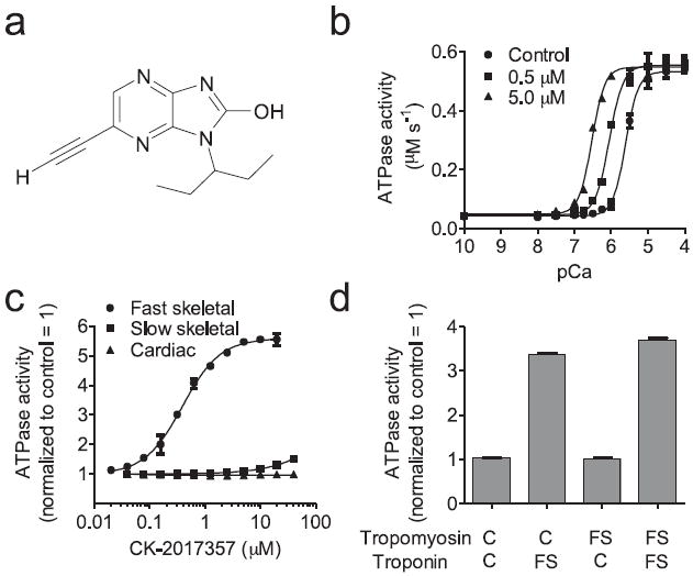
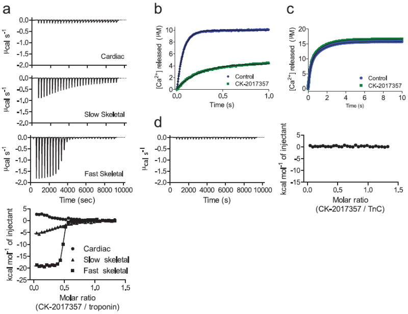
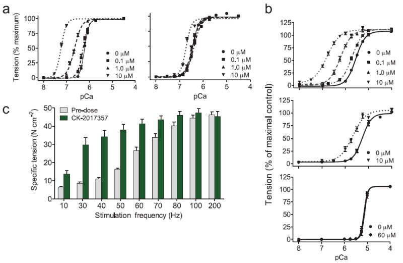
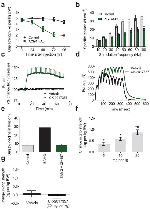
Limited neural input results in muscle weakness in neuromuscular disease because of a reduction in the density of muscle innervation, the rate of neuromuscular junction activation or the efficiency of synaptic transmission. We developed a small-molecule fast-skeletal-troponin activator, CK-2017357, as a means to increase muscle strength by amplifying the response of muscle when neural input is otherwise diminished secondary to neuromuscular disease. Binding selectively to the fast-skeletal-troponin complex, CK-2017357 slows the rate of calcium release from troponin C and sensitizes muscle to calcium. As a consequence, the force-calcium relationship of muscle fibers shifts leftwards, as does the force-frequency relationship of a nerve-muscle pair, so that CK-2017357 increases the production of muscle force in situ at sub-maximal nerve stimulation rates. Notably, we show that sensitization of the fast-skeletal-troponin complex to calcium improves muscle force and grip strength immediately after administration of single doses of CK-2017357 in a model of the neuromuscular disease myasthenia gravis. Troponin activation may provide a new therapeutic approach to improve physical activity in diseases where neuromuscular function is compromised.


Conflict of interest statement

COMPETING FINANCIAL INTERESTS AJR, JJH, ACH, ARM, RK, LD, GG, KHL, DM, WFB, MMC, DC, SEC, MG, RH, ZJ, PL, HR, KGS, JS, VV, DJM, BPM, FIM are past or present employees of Cytokinetics Inc. and own stock or stock options in Cytokinetics. DLA and DRC received research support from Cytokinetics, Inc.

Figures

FIGURE 1
FIGURE 1. CK-2017357 is a selective calcium sensitizer of the fast skeletal troponin complex
(a) The chemical structure of CK-2017357. (b) Calcium dependence of the fast skeletal myofibril ATPase at three concentrations of CK-2017357. (c) The dose response of CK-2017357 treated fast skeletal, slow skeletal and cardiac myofibrils at pCa 6.0. (d) Activation of heterologous, reconstituted thin filaments (source of regulatory complex isoform, C = cardiac, FS = fast skeletal) by CK-2017357 (20 μM) using cardiac myosin ATPase (mean ± SD) as a probe for thin filament activation at a pCa 6.75, approximately the pCa25 for all systems.
FIGURE 2
FIGURE 2. CK-2017357 binds to the skeletal troponin complex and slows calcium release
(a) Isothermal titration calorimetry of CK-2017357 added into purified cardiac, slow skeletal, and fast skeletal intact troponin complex (bottom panel). Shown above are representative experiments for the heats of addition to each isoform during the CK-2017357 titration. (b) Fluorescence intensity of the calcium chelator, Quin-2, following rapid mixing with intact fast skeletal troponin (10 μM final concentration) or (c) recombinant rabbit fast skeletal muscle troponin C (10 μM final concentration) in the presence or absence of CK-2017357 (20 μM final concentration). (d) Isothermal titration calorimetry of CK-2017357 added to recombinant rabbit fast skeletal troponin C (50 μM).
FIGURE 3
FIGURE 3. CK-2017357 shifts the force-calcium relationship in fast skeletal muscle leftwards and amplifies the response of muscle to nervous input
(a) The force-calcium relationship in human skinned fast (left) and slow (right) skeletal muscle fibers vs. calcium concentration at three concentrations of CK-2017357. Force measurements (each condition, n = 10) are displayed as a fraction of maximal force (Fo). (b) The force-calcium relationship of single skinned fibers from (top) rabbit fast skeletal psoas muscle fibers (n = 12), (middle) rat slow skeletal soleus muscle (n = 13), and (bottom) rat cardiac muscle fibers (n = 10) treated with CK-2017357. (c) The force-frequency relationship of rat EDL muscle in situ pre- and post-treatment with CK-2017357 (10 mg kg-1, administered into the gastrointestinal tract via a duodenal cannula to simulate oral administration, n = 6). For all graphs, mean ± SEM.
FIGURE 4
FIGURE 4. CK-2017357 improves muscle and physical function in a model of neuromuscular disease
(a) Rat forelimb grip strength in PT-EAMG rats vs. time after administration of nAChR antibody α1/3/5 or phosphate buffered saline (control), n = 10 each group. (b) The force-frequency relationship of rat EDL muscle in situ 72-96 hours after intra-peritoneal injection (500 μg kg-1) of nAChRα1/3/5 antibody (PT-EAMG, n = 6) or phosphate buffered saline (control, n = 4). (c) In situ rat EDL peak muscle force vs. time after infusion of CK-2017357 (3 mg kg-1, n = 6) or vehicle (n = 4) in PT-EAMG rats. (d) In situ rat EDL muscle force vs. time for a single representative contraction (10 stimuli at 30 Hz) before and after administration of CK-2017357 (3 mg kg-1) in PT-EAMG rats. (e) Decline in muscle force (sag) under the same stimulation protocol in control rats (n = 4), PT-EAMG rats (n = 6), or PT-EAMG rats after infusion of CK-2017357 (n = 6). (f) Forelimb grip strength in PT-EAMG rats 60 minutes following a single oral dose of CK-2017357 (n = 20-22 in each dose group. * P < 0.001 vs. vehicle, # P < 0.01 vs. lower doses). (g) Rat forelimb grip strength change after oral administration of CK-2017357 (n = 14) or vehicle (n = 14) in healthy rats. For all graphs, mean ± SEM plotted.

Comment in

References

    1. Classification of the neuromuscular disorders. Lancet. 1968;1:1020–1021. - PubMed
    1. Pollard T, Earnshaw W. Cell Biology. Saunders/Elsevier; Philadelphia: 2008.
    1. Freund HJ. Motor unit and muscle activity in voluntary motor control. Physiol Rev. 1983;63:387–436. - PubMed
    1. Jasmin BJ, Gardiner PF. Patterns of EMG activity of rat plantaris muscle during swimming and other locomotor activities. J Appl Physiol. 1987;63:713–718. - PubMed
    1. Sharma KR, Miller RG. Electrical and mechanical properties of skeletal muscle underlying increased fatigue in patients with amyotrophic lateral sclerosis. Muscle Nerve. 1996;19:1391–1400. - PubMed

Publication types

MeSH terms