

Paraganglioma of seminal vesicle and chromophobe renal cell carcinoma: a case report and literature review
- PMID: 22344361
- PMCID: PMC10906681
- DOI: 10.1590/s1516-31802012000100010
Paraganglioma of seminal vesicle and chromophobe renal cell carcinoma: a case report and literature review
Abstract
Context: Extra-adrenal paragangliomas are rare tumors that have been reported in many locations, including the kidney, urethra, urinary bladder, prostate, spermatic cord, gallbladder, uterus and vagina.
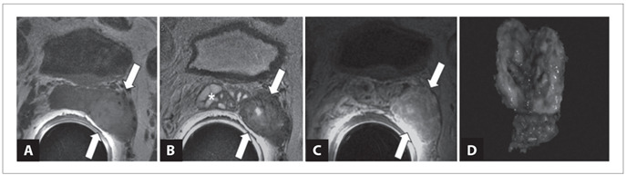
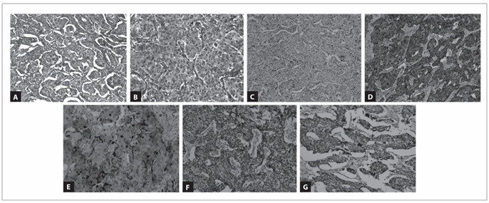
Case report: This report describes, for the first time to the best of our knowledge, a primary paraganglioma of the seminal vesicle occurring in a 61-year-old male. The patient presented persistent arterial hypertension and a previous diagnosis of chromophobe renal cell carcinoma. It was hypothesized that the seminal vesicle tumor could be a metastasis from the chromophobe renal cell carcinoma. Immunohistochemical characterization revealed expression of synaptophysin and chromogranin in tumor cell nests and peripheral S100 protein expression in sustentacular cells. Succinate dehydrogenase A and B-related (SDHA and SDHB) expression was present in both tumors.
Conclusions: No genetic alterations to the VHL and SDHB genes were detected in either the tumor tissue or tissues adjacent to the tumor, which led us to rule out a hereditary syndrome that could explain the association between paraganglioma and chromophobe renal cell carcinoma in a patient with arterial hypertension.
CONTEXTO:: Paragangliomas extra-adrenais são tumores raros que têm sido relatados em muitas localizações, incluindo rim, uretra, bexiga, próstata, cordão espermático, vesícula biliar, útero e vagina.
RELATO DE CASO:: Este relato descreve, pela primeira vez em nosso conhecimento, um paraganglioma primário da vesícula seminal ocorrendo em um paciente do sexo masculino de 61 anos de idade. O paciente apresentou hipertensão arterial persistente e um diagnóstico prévio de carcinoma de células renais cromófobo (CCRC). Foi pensado que o tumor de vesícula seminal poderia ser uma metástase do CCRC. A caracterização imunoistoquímica revelou expressão de sinaptofisina e cromogranina nos ninhos de células tumorais e expressão de proteína S100 nas células sustentaculares. Expressão de succinato de-hidrogenase A e B relacionada (SDHA e SDHB) estiveram presentes em ambos os tumores.
CONCLUSÕES:: Nenhuma alteração genética dos genes VHL e SDHB foi detectada nos tecidos tumorais e adjacentes ao tumor, o que nos levou a afastar uma síndrome hereditária que poderia explicar a associação entre o paraganglioma e o CCRC em um paciente com hipertensão arterial.
Conflict of interest statement
Figures
References
-
- Tischler AS. Pheochromocytoma and extra-adrenal paraganglioma: updates. Arch Pathol Lab Med. 2008;132(8):1272–1284. - PubMed
-
- Gupta R, Howell RS, Amin MB. Paratesticular paraganglioma: a rare cause of an intrascrotal mass. Arch Pathol Lab Med. 2009;133(5):811–813. - PubMed
-
- Lee JA, Duh QY. Sporadic paraganglioma. World J Surg. 2008;32(5):683–687. - PubMed
-
- Erickson D, Kudva YC, Ebersold MJ, et al. Benign paragangliomas: clinical presentation and treatment outcomes in 236 patients. J Clin Endocrinol Metab. 2001;86(11):5210–5216. - PubMed
-
- McNicol AM. Histopathology and immunohistochemistry of adrenal medullary tumors and paragangliomas. Endocr Pathol. 2006;17(4):329–336. - PubMed
Publication types
MeSH terms
Substances
LinkOut - more resources
Full Text Sources
Medical
Miscellaneous
