. 2012 Feb 15;4(121):121ra18.
doi: 10.1126/scitranslmed.3003205.

MicroRNA-21 promotes fibrosis of the kidney by silencing metabolic pathways

Affiliations

MicroRNA-21 promotes fibrosis of the kidney by silencing metabolic pathways

B Nelson Chau et al. Sci Transl Med. .

Abstract

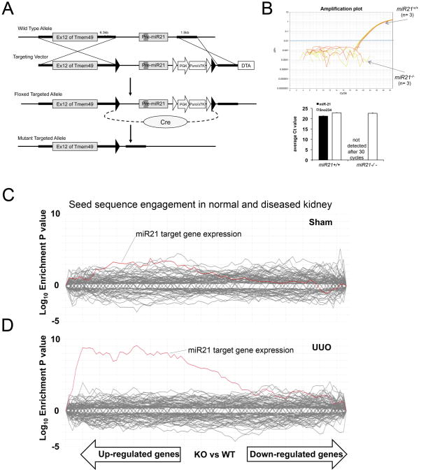
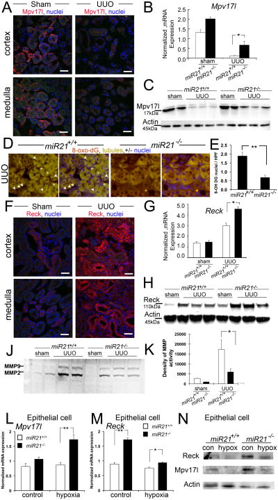
Scarring of the kidney is a major public health concern, directly promoting loss of kidney function. To understand the role of microRNA (miRNA) in the progression of kidney scarring in response to injury, we investigated changes in miRNA expression in two kidney fibrosis models and identified 24 commonly up-regulated miRNAs. Among them, miR-21 was highly elevated in both animal models and in human transplanted kidneys with nephropathy. Deletion of miR-21 in mice resulted in no overt abnormality. However, miR-21(-/-) mice suffered far less interstitial fibrosis in response to kidney injury, a phenotype duplicated in wild-type mice treated with anti-miR-21 oligonucleotides. Global derepression of miR-21 target mRNAs was readily detectable in miR-21(-/-) kidneys after injury. Analysis of gene expression profiles up-regulated in the absence of miR-21 identified groups of genes involved in metabolic pathways, including the lipid metabolism pathway regulated by peroxisome proliferator-activated receptor-α (Pparα), a direct miR-21 target. Overexpression of Pparα prevented ureteral obstruction-induced injury and fibrosis. Pparα deficiency abrogated the antifibrotic effect of anti-miR-21 oligonucleotides. miR-21 also regulated the redox metabolic pathway. The mitochondrial inhibitor of reactive oxygen species generation Mpv17l was repressed by miR-21, correlating closely with enhanced oxidative kidney damage. These studies demonstrate that miR-21 contributes to fibrogenesis and epithelial injury in the kidney in two mouse models and is a candidate target for antifibrotic therapies.


Conflict of interest statement

Competing interests: All authors from Regulus Therapeutics have stock options in the company. JSD is on the Scientific Advisory Board at Regulus Therapeutics and the Duffield Lab has a Sponsored Research Agreement with Regulus Therapeutics. JSD is on the Scientific Advisory Board at, and has stock options in, Promedior Inc.

Figures

Fig. 1
Fig. 1
MicroRNA 21 is upregulated in injury with fibrosis in mouse and human kidney. (A-B) Plots showing significantly upregulated microRNAs [red box, P < 0.001] detected on Agilent microRNA microarrays (A) after d10 of murine kidney UUO compared to normal kidney and (B) after 10d of murine unilateral kidney IRI compared with normal kidney. (C) Time course Q-PCR of whole kidney total RNA for miR-21 normalized to Sno234, from mice with UUO or IRI [** P < 0.01, * P< 0.05]. (D) RT-qPCR for miR-21 normalized to RNU19 in human kidney biopsies from patients with kidney transplantation (n= 5/group). Biopsies from healthy donor kidneys were normal or patients with CAD or transplant AKI was determined by histological assessment. (E) Q-PCR for miR-21 normalized to Sno234 from total RNA of purified endothelial cells, pericyte/myofibroblasts, macrophages and proximal epithelial cells from normal mouse kidney or d2 or d7 of UUO kidney. (F) Comparative Q-PCR from purified cells from normal kidney. (G) In situ hybridization of normal or post IRI murine kidney sections from miR-21+/+ or miR-21-/- mice for miR-21. Purple stain shows the presence of miR-21. Note in normal kidney some epithelium in medulla and papilla and some perivascular cells have miR-21 but in post IRI kidney expression of miR-21 is widespread in the kidney. [v = venule, a = arteriole, g = glomerulus, bar = 50μm] (n = 3-7/group. * P<0.05, **P<0.01).
Fig. 2
Fig. 2
Development and characterization of miR-21 targeted mutation in mice. (A) Schema showing targeting strategy for miR-21 in intron 12 of the Tmem49 gene. LoxP sites [black triangles] were inserted around Pre-miR-21 and the inserted puromycin resistance gene. Cre mediated excision was performed in ES cells prior to generation of mutant mice. (B) Taqman Amplification plot (upper) showing amplification of miR-21 in RNA extracted from miR21+/+ kidney but not miR21-/- kidney, and graph (lower) showing expression level of mature miR-21 from miR-21+/+ and miR-21-/- kidneys. The small nucleolar RNA Sno234 was used as a control. (C-D) SylArray analyses (16) showing whole kidney microarray data [30,000 genes] from miR-21-/- compared with miR-21+/+ kidneys. Each line shows the relative expression intensity [miR-21-/- relative to miR-21+/+] for all mRNA transcripts that contain a target (seed) sequence for a particular microRNA in their 3′ untranslated region. The red line identifies all mRNA transcripts with seed sequence for miR-21. In normal miR-21-/- kidney (C), there is no significant enrichment of miR-21 target genes, but in miR-21-/- kidney d7 after UUO (D) there is marked and highly significant enrichment of expression of miR-21 target gene transcripts in the microarray.
Fig. 3
Fig. 3
Gene targeted mutation indicates that microRNA 21 amplifies injury and fibrosis in the kidney. (A) Panels showing low power images of d10 UUO injured or sham treated kidneys from miR-21-/- or miR-21+/+ matched mice. (B) Quantification of Sirius red stained fibrosis in kidneys. (C) Q-PCR quantification of Coll1a1 and Coll3a1 transcripts normalized to Gapdh. (D) Number of apoptotic cells per high power field on TUNEL stained kidney sections. (E) Epithelia injury scores; either number of epithelial cells with brush border or number of tubules with cells containing brush border. (F) Western Blot showing Kim1 protein levels in miR-21-/- or miR-21+/+ UUO kidneys. (G-J) Morphometric quantification of (G) αSMA stained myofibroblasts (H) endothelial density (J) macrophages. (K) Panels showing low power images of d10 post IRI or sham treated kidneys from miR-21-/- or miR-21+/+ matched mice. (L) Quantification of Sirius red stained fibrosis in kidneys. (M) Q-PCR quantification of Coll1a1 and Coll3a1 transcripts normalized to Gapdh (n = 6-9/group. * P<0.05, **P<0.01 ***P<0.001. Bar = 100μm).
Fig. 4
Fig. 4
Silencing microRNA 21 in vivo ameliorates fibrosis and albuminuria in the kidney. (A) Panels of normal and diseased kidney showing detection of Cy3 tagged anti-miR given by a single IP injection (20mg/kg) 7 days previously, and co-labeled for pericytes/myofibroblasts with anti-Pdgfrβ antibodies (green). Note intense red color in epithelium but also in vascular and perivascular cells (arrowheads) [g=glomerulus]. Prevention Study (panels B-F): (B-C) Low power images (B) and morphometric quantification (C) of sirius red stained kidneys for fibrosis at d10 of UUO model, in wild-type mice given anti-miR-21 or a control anti-miR by IP injection. (D) Q-PCR results for pathological collagen transcripts normalized to Gapdh in the UUO model. (E-F) Morphometric quantification (E) and Q-PCR (F) for collagen transcripts in kidneys subjected to unilateral IRI and given anti-miR-21 or a control anti-miR by IP injection. Reversal Study (panels G-K): (G-J) Fibrosis and collagen transcripts in mice subjected to unilateral IRI 5 days prior to delivery of anti-miR-21 or a control anti-miR by IP injection, and assessed at d14 after IRI. (K) Urinary Albumin:Creatinine ratio in mice after unilateral IRI and given anti-miR-21 or a control anti-miR by IP injection, followed at 7 days by removal of the healthy kidney and urine assessed at one day later (n= 7-16/group. * P<0.05, **P<0.01. Bar = 100μm).
Fig. 5
Fig. 5
The lipid metabolism pathway and Pparα are miR-21 targets in kidney injury. (A) De-repressed genes from microarray analysis of UUO kidneys from miR-21-/- or miR-21+/+ mice by biological process. Green bars highlight lipid metabolism (B) Heat map (blue-low, red, high) comparing lipid metabolism genes that are over-represented in Pparα transgenic mouse muscle with miR-21 deficient UUO kidneys (C) Photomicrogaphs showing expression of Pparα in normal and UUO kidney. (D) Q-PCR for Ppara transcripts normalized to Gapdh in miR-21-/- or miR-21+/+ mice. (E-F) Western blot (F) and band intensity quantification (E) for Pparα in UUO kidneys from miR-21-/- or miR-21+/+ mice. (G-H) Q-PCR and Western blot of confluent monolayers of primary epithelial cultures for Pparα from kidneys from miR-21-/- or miR-21+/+ mice, in normal culture conditions or following 24h of hypoxia. Schema (J) and characterization (K-N) of the response of kidneys to UUO injury during over-expression of Pparα in androgen sensitive conditional KAP2-Ppara transgenic female mice. (K) Ppara expression in normal kidneys 5d after implantation of testosterone (t) or sham. (L) Q-PCR quantification of Mcad transcripts, (M) Western Blot and quantification of αSMA in sham or UUO kidneys of Kap2-Ppara or WT mice treated with testosterone and (N) Quantification of Sirius red stained fibrosis. (P-R) Characterization of the response of Ppara-/- kidneys to anti-miR21 administration. Graphs quantifying Sirius red stained fibrosis (P) and Collagen transcript quantification in UUO or sham surgery kidneys (Q), or epithelial injury scores (R) in UUO kidneys. (n= 3-7/group. * P<0.05, **P<0.01. Bar = 100μm).
Fig. 6
Fig. 6
The mitochondrial redox regulator Mpv17-like and the metalloproteinase inhibitor Reck are miR-21 targets in kidney injury and are important endogenous inhibitors of injury and fibrosis. (A) Photomicrographs showing expression of Mpv17-like. (B) Q-PCR for Mpv17l transcripts normalized to Gapdh in miR-21-/- or miR-21+/+ matched mice. (C) Western blot Mpv17l in UUO kidneys from miR-21-/- or miR-21+/+ matched mice. (D-E) Images (D) and quantification (E) of ROS production as detected by the oxidized derivative of deoxyguanosine in nuclei of kidney cells in miR-21-/- or miR-21+/+matched mice. (F) Photomicrographs showing expression of Reck. (G-H) Q-PCR for Reck transcripts normalized to Gapdh (G) Western blot for Reck (H) in normal or UUO kidneys from miR-21-/- or miR-21+/+ matched mice. (J-K) Gelatin zymography (J) and band density quantification (K) of whole cell lysates from UUO kidneys from miR-21-/- or miR-21+/+ matched mice. (L-N) Hypoxia studies on primary kidney epithelial cells cultures showing Q-PCR (L-M) and Western blots (N) for Mpv17l and Reck. (n= 3-7/group. * P<0.05, **P<0.01. Bar = 100μm).

References

    1. Lupher ML, Jr, Gallatin WM. Regulation of fibrosis by the immune system. Adv Immunol. 2006;89:245–288. - PubMed
    1. Hernandez-Gea V, Friedman SL. Pathogenesis of liver fibrosis. Annu Rev Pathol. 6:425–456. - PubMed
    1. Castano AP, et al. Serum amyloid P inhibits fibrosis through Fc gamma R-dependent monocyte-macrophage regulation in vivo. Sci Transl Med. 2009;1:5ra13. - PMC - PubMed
    1. Lin SL, et al. Targeting endothelium-pericyte cross talk by inhibiting VEGF receptor signaling attenuates kidney microvascular rarefaction and fibrosis. Am J Pathol. 2011;178:911–923. - PMC - PubMed
    1. Thum T, et al. MicroRNA-21 contributes to myocardial disease by stimulating MAP kinase signalling in fibroblasts. Nature. 2008;456:980–984. - PubMed

Publication types