. 2012 May;19(5):483-92.
doi: 10.1177/1933719111432870. Epub 2012 Feb 16.

HDAC1 and HDAC2 are differentially expressed in endometriosis

Affiliations

HDAC1 and HDAC2 are differentially expressed in endometriosis

Maricarmen Colón-Díaz et al. Reprod Sci. 2012 May.

Abstract

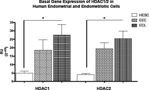
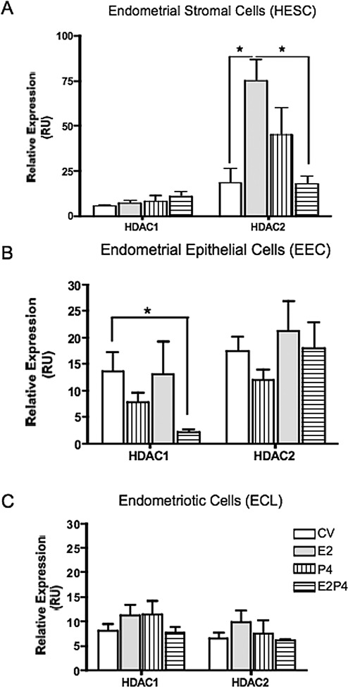
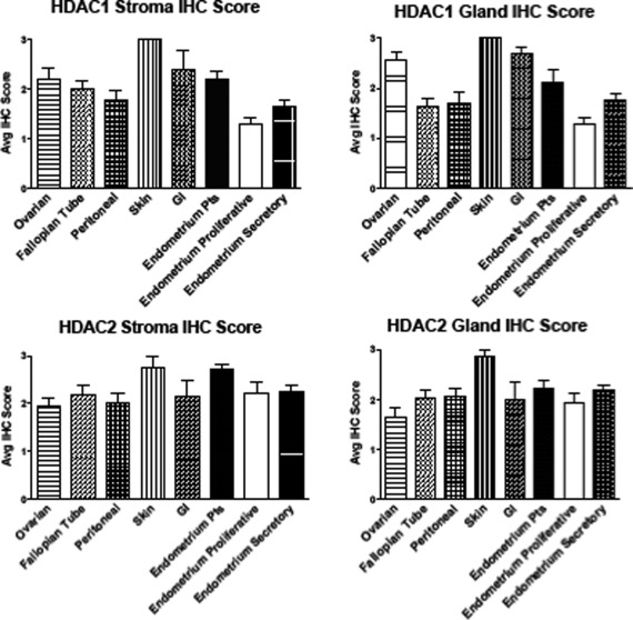
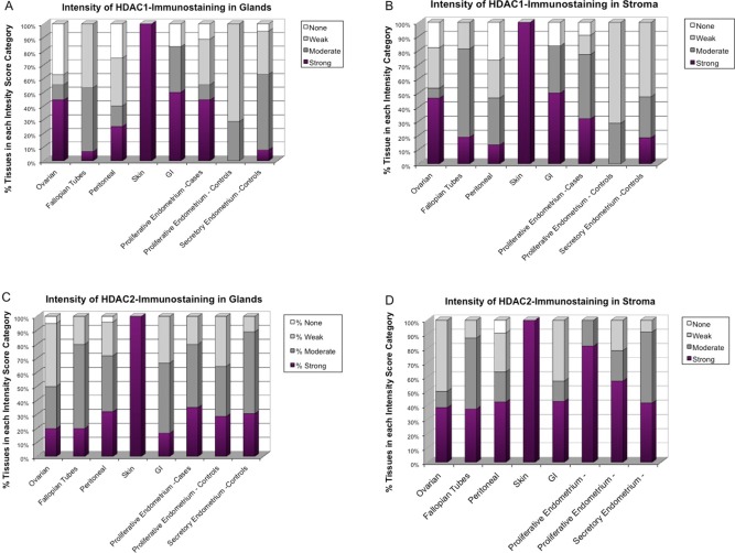
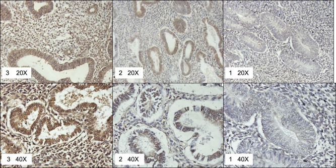
Epigenetic mechanisms have been ascribed important roles in endometriosis. Covalent histone modifications at lysine residues have been shown to regulate gene expression and thus contribute to pathological states in many diseases. In endometriosis, histone deacetylase inhibition (HDACi) resulted in reactivation of E-cadherin, attenuation of invasion, decreased proliferation of endometriotic cells, and caused lesion regression in an animal model. This study was conducted to assess basal and hormone-regulated gene expression levels of HDAC1 and HDAC2 (HDAC1/2) in cell lines and protein expression levels in tissues. Basal and steroid hormone-regulated HDAC1/2 gene expression levels were determined by quantitative polymerase chain reaction in cell lines and tissues. Protein levels were measured by immunohistochemistry (IHC) in tissues on an endometriosis tissue microarray (TMA). Basal HDAC1/2 gene expression levels were significantly higher in endometriotic versus endometrial stromal cells, which was confirmed by Western blot analysis. Estradiol (E2) and progesterone (P4) significantly downregulated HDAC1 expression in endometrial epithelial cells. Levels of HDAC2 were upregulated by E2 and downregulated by E2 + P4 in endometrial stromal cells. Hormone modulation of HDAC1/2 gene expression was lost in the endometriotic cell line. Immunohistochemistry showed that HDAC1/2 proteins were expressed in a substantial proportion of lesions and endometrium from patients, and their expression levels varied according to lesion localization. The highest proportion of strong HDAC1 immunostaining was seen in ovarian, skin, and gastrointestinal lesions, and of HDAC2 in skin lesions and endometrium from patients with endometriosis. These studies suggest that endometriosis etiology may be partially explained by epigenetic regulation of gene expression due to dysregulations in the expression of HDACs.


Conflict of interest statement

The authors declared no potential conflicts of interest with respect to the research, authorship, and/or publication of this article.

Figures

Figure 1.
Figure 1.
Basal gene expression of class I histone deacetylases (HDACs) in human endometrial and endometriotic cell lines. Quantitative polymerase chain reaction (qPCR) quantification of class I HDACs (HDAC1 and HDAC2) was conducted using TaqMan gene expression assays. Results are expressed as mean ± standard deviation (SD) of at least 3 experiments in duplicate. Both HDAC1 and HDAC2 are differentially expressed in human endometrial cell lines. Endometriotic cells express significantly higher levels of HDAC1/2. HESC, endometrial stromal; EEC, endometrial epithelial; ECL, endometriotic cell line. *P < .05.
Figure 2.
Figure 2.
Effects of ovarian steroid hormone treatment on histone deacetylase (HDAC) 1/2 gene expression in human endometrial and endometriotic cell lines. Cells were treated for 24 hours with 10−8 mol/L estradiol (E2), 10−7 mol/L progesterone (P4), or E2/P4 for 24 hours. Total RNA was isolated from treated and untreated (medium with vehicle, CV) cells. Quantitative polymerase chain reaction (qPCR) of HDAC1/2 was conducted using TaqMan gene expression assays. Results are expressed as relative expression (relative units, RU) after normalization with the reference gene. The HDAC1 gene expression was significantly downregulated by E2/P4 in endometrial epithelial cells (B) but not in endometrial stromal nor in the endometriotic cells (C). The HDAC2 gene expression levels were significantly increased by E2 and dowregulated by E2 + P4 in endometrial stromal cells (A). **P < .01.
Figure 3.
Figure 3.
Gene expression levels of histone deacetylase (HDAC) 1/2 in endometrial and endometriotic tissues. A, Relative gene expression levels in tissues: RNA was isolated from flash-frozen tissues and quantitative polymerase chain reaction (qPCR) was conducted using TaqMan assays. Results are expressed as relative units (RU) after normalization with the reference gene glyceraldehyde 3-phosphate dehydrogenase (GAPDH). The HDAC1 was expressed in tissues of patients with endometriosis (endometriotic and eutopic endometrium), and in the endometria of patients with fibroids. The highest mean expression of HDAC1 was observed in the endometria of patients with endometriosis. Similar results were observed for HDAC2, which was expressed in a substantial proportion of tissues from patients with endometriosis and fibroids. The highest expression of HDAC2 was in endometria from patients with endometriosis. B, Correlations of HDAC1/2 gene expression levels in tissues: a positive and significant correlation was observed in the levels of HDAC1/2 only in endometriotic lesions.
Figure 4.
Figure 4.
Immunostaining intensity of histone deacetylase (HDAC)1/2 proteins in endometrial and endometriotic tissues. A total of 164 formalin-fixed paraffin-embedded endometrial and endometriotic tissues on a tissue microarray (TMA) were analyzed by immunohistochemistry. Immunostaining intensity of HDAC1 and HDAC2 was evaluated and scored blindly in glands (A and C) and stroma (B and D) by 2 independent observers using the intensity scale (3 = strong, 2 = moderate, 1 = weak, and 0 = no staining). Staining was reported as mean immunohistochemistry (IHC) score ± standard deviation (SD). The highest mean expression of HDAC1 was seen in skin lesions followed by gastrointestinal (GI), ovarian, and endometrium from patients with endometriosis (EE). Differences in mean HDAC2 immunostaining intensity were subtler, with the highest mean expression seen in skin, EE, and GI lesions. The level of expression of HDAC2 in EE was significantly higher than secretory phase endometrium (SE).
Figure 5.
Figure 5.
Proportion of tissues in each histone deacetylase (HDAC)1/2 immunostaining intensity category in endometrial and endometriotic tissues. The proportion of tissues in each tissue type categorized as having strong (3), moderate (2), weak (1), or no (0) immunostaining for HDAC1 and HDAC2 was determined. At the protein level, HDAC1/2 were differentially expressed according to lesion localization, with the highest proportion of positive HDAC1 immunostaining seen in ovarian, skin, and gastrointestinal (GI) lesions, and of HDAC2 in skin and endometria of patients with endometriosis.
Figure 6.
Figure 6.
Representative pictures of histone deacetylase (HDAC)1-specific intensities scored as strong (3), moderate (2), or weak (1) on the tissue microarray (TMA). ×20 and ×40 pictures are provided for detail.

References

    1. Bulun SE. Endometriosis. N Engl J Med. 2009;360(3):268–279 - PubMed
    1. Giudice LC, Kao LC. Endometriosis. Lancet. 2004;364(9447):1789–1799 - PubMed
    1. Flores I, Rivera E, Ruiz LA, Santiago OI, Vernon MW, Appleyard CB. Molecular profiling of experimental endometriosis identified gene expression patterns in common with human disease. Fertil Steril. 2007;87(5):1180–1199 - PMC - PubMed
    1. Konno R, Fujiwara H, Netsu S, et al. Gene expression profiling of the rat endometriosis model. Am J Reprod Immunol. 2007;58(4):330–343 - PubMed
    1. Eyster KM, Boles AL, Brannian JD, Hansen KA. DNA microarray analysis of gene expression markers of endometriosis. Fertil Steril. 2002;77(1):38–42 - PubMed

Publication types

MeSH terms