Review
. 2012 Mar;50(3):164-75.
doi: 10.1002/dvg.22007. Epub 2012 Feb 16.

The hitchhiker's guide to Xenopus genetics

Affiliations
Review

The hitchhiker's guide to Xenopus genetics

Anita Abu-Daya et al. Genesis. 2012 Mar.

Abstract

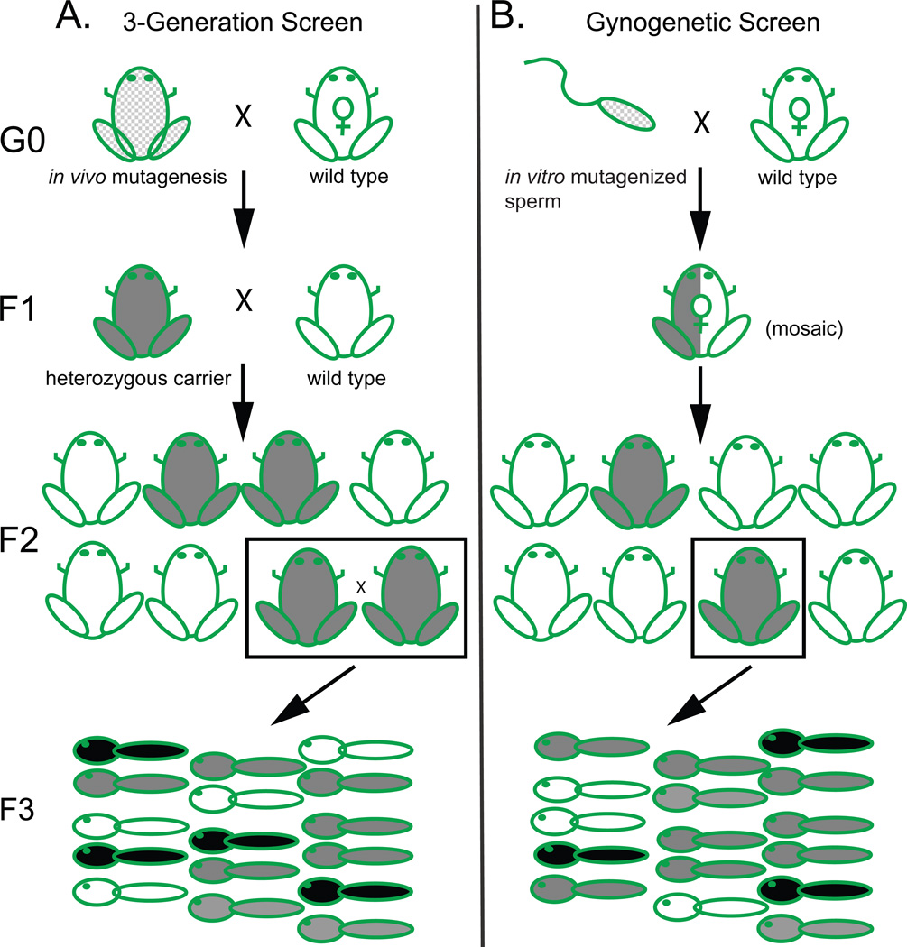
A decade after the human genome sequence, most vertebrate gene functions remain poorly understood, limiting benefits to human health from rapidly advancing genomic technologies. Systematic in vivo functional analysis is ideally suited to the experimentally accessible Xenopus embryo, which combines embryological accessibility with a broad range of transgenic, biochemical, and gain-of-function assays. The diploid X. tropicalis adds loss-of-function genetics and enhanced genomics to this repertoire. In the last decade, diverse phenotypes have been recovered from genetic screens, mutations have been cloned, and reverse genetics in the form of TILLING and targeted gene editing have been established. Simple haploid genetics and gynogenesis and the very large number of embryos produced streamline screening and mapping. Improved genomic resources and the revolution in high-throughput sequencing are transforming mutation cloning and reverse genetic approaches. The combination of loss-of-function mutant backgrounds with the diverse array of conventional Xenopus assays offers a uniquely flexible platform for analysis of gene function in vertebrate development.


Figures

Figure 1
Figure 1. Screen strategies
(A) (left column) Conventional 3-generation mutagenesis and screen scheme. In vivo mutagenesis of adult G0 males followed by successive outcrosses gives groups of F2 siblings, half carrying a given recessive allele (grey frogs). ¼ of random F2 sibling matings uncover the phenotype (black tadpoles) in 25% of their offspring. (B) (right column) Gynogenetic screen scheme. Dissociated testes are mutagenized in vitro and used to fertilize eggs, producing F1 founders mosaic for induced mutations (half-grey frog), which are then outcrossed to produce non-mosaic F2 carriers (grey). Gynogenesis from individual F2 carrier females uncovers recessive phenotypes (black tadpoles) in non-mendelian ratios of ~10–50% depending on gene-centromere distance.
Figure 2
Figure 2. Haploid genetics and gynogenesis
(A) As in normal oogenesis, recombination in meiosis I between non-sister chromatids from blue and red parental strains produces (B) oocytes arrested in meiosis II containing both sister chromatids from one parental strain, plus the 1st polar body. (C) To create gynogenetic haploids, sperm are UV-irradiated to block paternal genetic contribution, with the resulting zygote (D) completing second meiosis by ejecting the second set of sister chromatids in the 2nd polar body to give a haploid (E, Hoechst-stained karyotype below). Haploids can be rescued by allowing DNA replication to take place, then blocking cytokinesis with cold shock to form completely homozygous (isogenic) double haploids. Alternatively, (G) cold shock immediately after fertilization with UV-irradiated sperm suppresses polar body formation to create ‘early cold shock’ gynogenetic diploids retaining both sets of sister chromatids. Due to recombination (A), gynogenotes are not homozygous at all loci, but rescue to viable diploidy is very efficient.
Figure 3
Figure 3. High throughput mutation mapping strategies
(A) ‘red’ strain frog showing two chromosomes, one of which carries a mutation (*), crossed to a polymorphic ‘blue’ strain animal differing at many sequence loci. (B) Both parental chromosomes are inherited by hybrid ‘map cross’ offspring, and recombine in meiosis. (C) Gynogenetic diploids with recombined sister chromatid pairs are obtained from mapcross females and sorted into phenotypically mutant and wild type pools. In the homozygous mutant pool, red strain alleles dominate near the mutant locus, with increasing frequency of blue alleles further from the mutation; wild type usually show both alleles. (D) Recessive phenotypes mapping close to centromeres appear in up to 50% of gynogenotes, with frequency of wild type heterozygotes increasing with crossovers for more distal loci, providing gene-centromere distance. For all but very distal mutations, linkage to one of the 10 X. tropicalis centromeres can be detected. (E) A genomic region can then be inspected for candidate genes to evaluate (F). (G–K) Alternatively, mutations can be mapped or directly identified by whole-exome sequencing of carrier DNA from a toe clip. Pools of mutant and wild type embryo DNA are enriched for coding sequences by hybridization with synthetic biotinylated RNA baits and sequenced. (J) Sequences are then bioinformatically inspected for homozygous SNPs in the mutant pool to identify candidate genes or define a linked interval.
Figure 4
Figure 4. Selected X. tropicalis phenotypes
(A–D) The xenopus de milo (xdm) mutation reveals an unexpected requirement for nephronectin in limb subtype initiation upstream of the earliest known forelimb marker, tbx5; (C) WISH showing tbx5 forelimb field expression (arrow) and loss in mutant (D, arrow). (E–H) Heartbeat is absent in the cardiac myosin myh6 mutation muzak (muz) stained for sarcomeric myosin (green) and phalloidin (red). Despite absence of beating/contractility, heart looping and chamber formation are relatively normal, but (G,H) reconstruction from serial sections shows the endocardium has collapsed. (I,J) mutations affecting muscle structure (mrs lot (mlo)) disrupt birefringence to polarized light. (K,L) myosin chaperone unc45b mutant dicky ticker (dit) shows disordered sarcomere ultrastructure. (M–P) Mutations affecting inner ear development and the formation of otoconial crystals include seasick (ssk), in the vesicle transport adaptor protein ap3δ1, and komimi (kom), in the oc90 gene, as well as bunny (bun) where the otic vesicle is reduced in size. (Q–T) Multiciliate epidermal cells are affected in grinch (gri), with stubby cilia reduced in length and number in the mutant as seen stained with α-acetylated tubulin (Q,R) and by SEM (S,T).

References

    1. Abu-Daya A, Nishimoto S, Fairclough L, Mohun TJ, Logan MP, Zimmerman LB. The secreted integrin ligand nephronectin is necessary for forelimb formation in Xenopus tropicalis. Dev Biol. 2010;349:204–212. - PMC - PubMed
    1. Abu-Daya A, Sater AK, Wells DE, Mohun TJ, Zimmerman LB. Absence of heartbeat in the Xenopus tropicalis mutation muzak is caused by a nonsense mutation in cardiac myosin myh6. Developmental biology. 2009;336:20–29. - PMC - PubMed
    1. Adams D. Life, The Universe and Everything. 1982
    1. Amaya E, Musci TJ, Kirschner MW. Expression of a dominant negative mutant of the FGF receptor disrupts mesoderm formation in Xenopus embryos. Cell. 1991;66:257–270. - PubMed
    1. Bai X, Kim J, Yang Z, Jurynec MJ, Akie TE, Lee J, LeBlanc J, Sessa A, Jiang H, DiBiase A, Zhou Y, Grunwald DJ, Lin S, Cantor AB, Orkin SH, Zon LI. TIF1gamma controls erythroid cell fate by regulating transcription elongation. Cell. 2010;142:133–143. - PMC - PubMed

Publication types

LinkOut - more resources