. 2012 May;112(9):1564-75.
doi: 10.1152/japplphysiol.00027.2012. Epub 2012 Feb 16.

Akt and MAPK signaling mediate pregnancy-induced cardiac adaptation

Affiliations

Akt and MAPK signaling mediate pregnancy-induced cardiac adaptation

Eunhee Chung et al. J Appl Physiol (1985). 2012 May.

Abstract

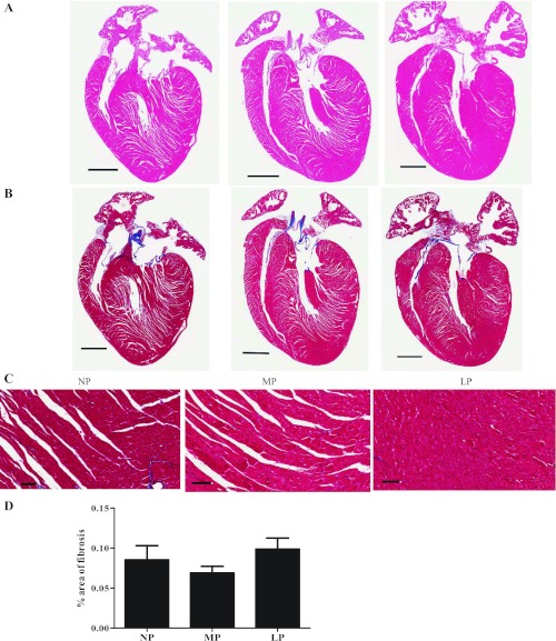
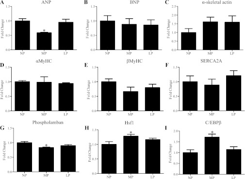
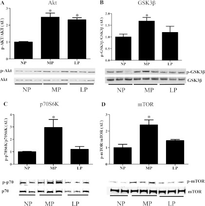
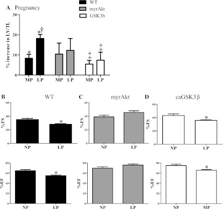
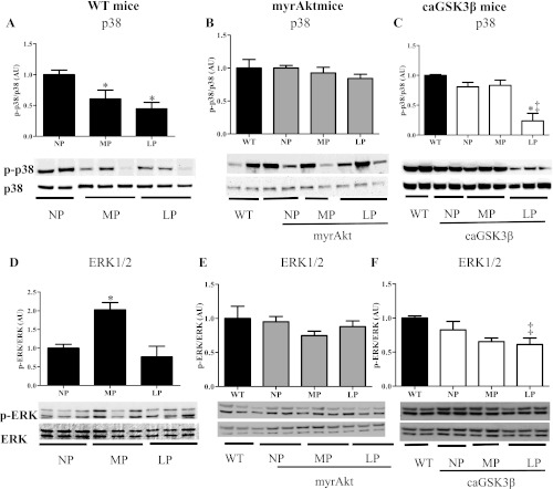
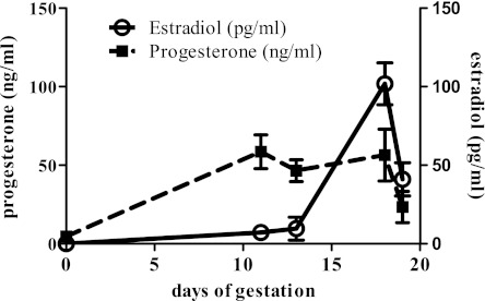
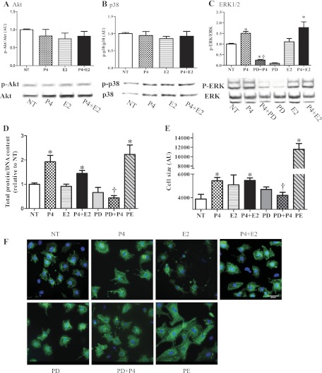
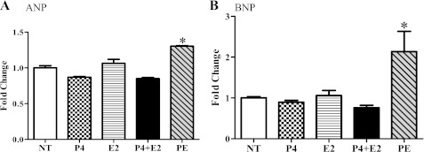
Although the signaling pathways underlying exercise-induced cardiac adaptation have been extensively studied, little is known about the molecular mechanisms that result in the response of the heart to pregnancy. The objective of this study was to define the morphological, functional, and gene expression patterns that define the hearts of pregnant mice, and to identify the signaling pathways that mediate this response. Mice were divided into three groups: nonpregnant diestrus control, midpregnancy, and late pregnancy. Both time points of pregnancy were associated with significant cardiac hypertrophy. The prosurvival signaling cascades of Akt and ERK1/2 were activated in the hearts of pregnant mice, while the stress kinase, p38, was decreased. Given the activation of Akt in pregnancy and its known role in cardiac hypertrophy, the hypertrophic response to pregnancy was tested in mice expressing a cardiac-specific activated (myristoylated) form of Akt (myrAkt) or a cardiac-specific constitutively active (antipathologic hypertrophic) form of its downstream target, glycogen synthase kinase 3β (caGSK3β). The pregnancy-induced hypertrophic responses of hearts from these mice were significantly attenuated. Finally, we tested whether pregnancy-associated sex hormones could induce hypertrophy and alter signaling pathways in isolated neonatal rat ventricular myocytes (NRVMs). In fact, progesterone, but not estradiol treatment increased NRVM cell size via phosphorylation of ERK1/2. Inhibition of MEK1 effectively blocked progesterone-induced cellular hypertrophy. Taken together, our study demonstrates that pregnancy-induced cardiac hypertrophy is mediated by activation of Akt and ERK1/2 pathways.


Figures

Fig. 1.
Fig. 1.
A: histological analysis of nonpregnancy (NP), midpregnancy (MP), and late pregnancy (LP) heart sections stained with hematoxylin and eosin (H and E). B: Masson's trichrome staining indicated that fibrosis was not induced during pregnancy. Scale bar, 0.5 mm. C: higher magnification (20×) of image in B. Scale bar, 0.625 mm. D: bar graph is average of % area of fibrosis from 3 hearts per group. Values are means ± SE.
Fig. 2.
Fig. 2.
qRT-PCR of selected genes in hearts of wild-type (WT) mice. Values are means ± SE expressed as fold change relative to nonpregnant control group (NP). qRT-PCR was performed in triplicate with a minimum of 4 independent left ventricular samples at different time points. The levels of all candidate genes were normalized to 18S rRNA. See Glossary for definition of abbreviations in A–I. *P < 0.05, significantly different from NP.
Fig. 3.
Fig. 3.
Pregnancy modulates Akt and its downstream targets in WT mice. A: Akt phosphorylation was significantly increased during MP and LP. B: GSK3β phosphorylation was significantly increased in MP. C: p70S6K phosphorylation was significantly increased in MP. D: mTOR phosphorylation was significantly increased in MP. Five to six animals were used per group with 2 to 3 technical replicates per animal, and a representative blot was shown. *P ≤ 0.05, significantly different from NP.
Fig. 4.
Fig. 4.
Cardiac mass adaptation in response to pregnancy of WT, myrAkt, and caGSK3β mice. A: in WT, the percent increases in left ventricular mass (LV) normalized to tibial length (LV/TL) were 8.3% and 18.2% in MP (n = 12) and LP (n = 18), respectively, compared with NP (n = 13). In myrAkt mice, LV/TL was increased 10.4% and 15.2% in MP (n = 8) and LP (n = 8), respectively, compared with NP (n = 8). In caGSK3β mice, LV/TL was increased 5.4% and 7.4% in MP (n = 9) and LP (n = 9), respectively, compared with NP (n = 17). B: %fractional shortening (%FS) and %ejection fraction (%EF) measured by echocardiography were significantly decreased in LP (n = 9 for NP and n = 8 for LP) in WT mice. C: %FS and %EF were maintained in LP in myrAkt mice (n = 11/group). D: %FS and %EF were significantly decreased in LP in caGSK3β mice (n = 5/group). Values are means ± SE. For abbreviations, see Glossary. *P < 0.05, significantly different from NP; †P < 0.05, significantly different from MP; ‡P < 0.05, significantly different from WT.
Fig. 5.
Fig. 5.
Pregnancy modulates MAPK signaling in hearts of WT and GSK3β mice, but not in hearts of myrAkt. p38 phosphorylation was significantly decreased during MP and LP in WT mice (A) but not in myrAkt (B). p38 phosphorylation was significantly decreased in LP in caGSK3β mice (C). ERK1/2 phosphorylation was significantly increased in MP but returned to NP levels in WT mice (D), but no alterations were observed in myrAkt mice (E); alterations were observed in LP caGSK3β mice (F). Five to six animals were used per group with 2 to 3 technical replicates per animal, and a representative blot was shown. *P ≤ 0.05, significantly different from NP; ‡P < 0.05, significantly different from WT.
Fig. 6.
Fig. 6.
Hormonal changes during pregnancy. Serum estradiol (E2) levels were below 10 pg/ml from NP to MP and peaked at LP and declined with approaching parturition. Serum progesterone levels were significantly increased at MP, maintained until approaching parturition.
Fig. 7.
Fig. 7.
The pregnancy hormone, progesterone (P4), causes neonatal rat ventricular myocyte (NRVM) hypertrophy via ERK signaling pathways. NRVMs treated with 300 nM P4, 500 pM estradiol (E2), or combinations of progesterone and estradiol (P4 + E2) did not show a change in Akt phosphorylation (A) or p38 phosphorylation (B), but P4 increased ERK1/2 phosphorylation (C). PD98059 (PD) blocked progesterone-induced ERK1/2 phosphorylation. We used triplicate wells from the same cell isolation for each condition, and data are the means ± SE of 6 independent experiments. D: total cellular protein normalized to DNA content was significantly increased by P4 and P4 + E2, but PD abolished the progesterone-induced increase in total protein concentration; 20 μM phenylephrine (PE) was used as a positive control. E: cell size was significantly increased by P4, P4 + E2, and PE treatment. P4-mediated cardiomyocyte hypertrophy was blocked by PD treatment. F: NRVMs were stained with α-actinin and cell size was measured. Representative images of 3 independent experiments are shown; scale bar is 20 μm. *P ≤ 0.05, significantly different from nontreated serum free group (NT); †P ≤ 0.05, significantly different from P4-treated group.
Fig. 8.
Fig. 8.
Pregnancy hormones did not change expression of fetal genes in NRVMs. A and B: mRNA levels measured by qRT-PCR. ANP (A) and BNP (B) were not altered with pregnancy hormones. PE was used as a positive control. *P ≤ 0.05, significantly different from nontreated serum free group (NT).

References

    1. Antos CL, McKinsey TA, Frey N, Kutschke W, McAnally J, Shelton JM, Richardson JA, Hill JA, Olson EN. Activated glycogen synthase-3beta suppresses cardiac hypertrophy in vivo. Proc Natl Acad Sci USA 99: 907–912, 2002 - PMC - PubMed
    1. Barnard RJ, Duncan HW, Baldwin KM, Grimditch G, Buckberg GD. Effects of intensive exercise training on myocardial performance and coronary blood flow. J Appl Physiol 49: 444–449, 1980 - PubMed
    1. Bocalini D, Carvalho E, de Sousa A, Levy R, Tucci P. Exercise training-induced enhancement in myocardial mechanics is lost after 2 weeks of detraining in rats. Eur J Appl Physiol 109: 909–914, 2010 - PubMed
    1. Boström P, Mann N, Wu J, Quintero PA, Plovie ER, Panáková D, Gupta RK, Xiao C, MacRae CA, Rosenzweig A, Spiegelman BM. C/EBP[beta] controls exercise-induced cardiac growth and protects against pathological cardiac remodeling. Cell 143: 1072–1083, 2010 - PMC - PubMed
    1. Bueno OF, De Windt LJ, Tymitz KM, Witt SA, Kimball TR, Klevitsky R, Hewett TE, Jones SP, Lefer DJ, Peng CF, Kitsis RN, Molkentin JD. The MEK1-ERK1/2 signaling pathway promotes compensated cardiac hypertrophy in transgenic mice. EMBO J 19: 6341–6350, 2000 - PMC - PubMed

Publication types

MeSH terms

LinkOut - more resources