. 2012 May;86(9):4743-51.
doi: 10.1128/JVI.06501-11. Epub 2012 Feb 15.

Hsp70 protein positively regulates rabies virus infection

Affiliations

Hsp70 protein positively regulates rabies virus infection

Xavier Lahaye et al. J Virol. 2012 May.

Abstract

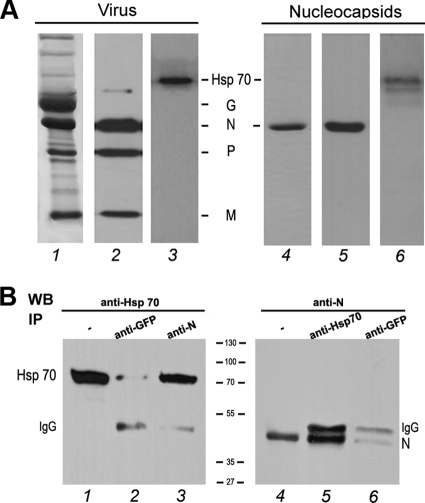
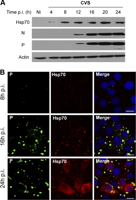
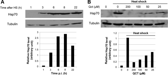
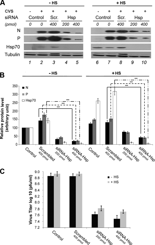
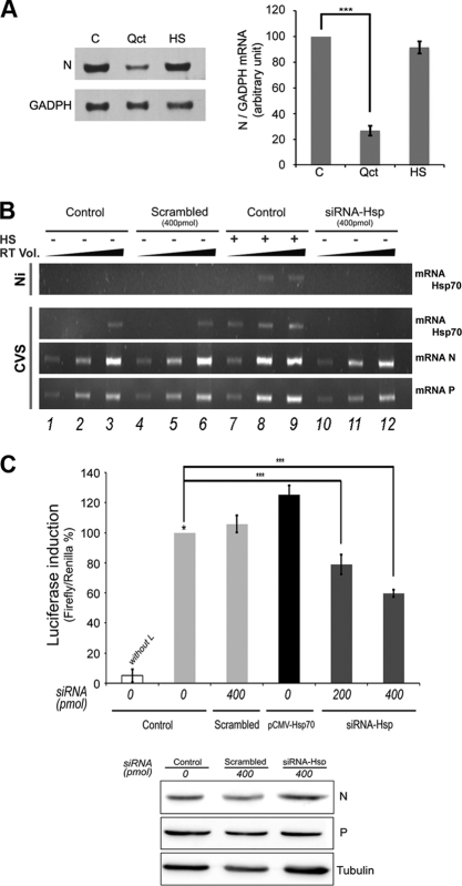
The Hsp70 chaperone plays a central role in multiple processes within cells, including protein translation, folding, intracellular trafficking, and degradation. This protein is implicated in the replication of numerous viruses. We have shown that rabies virus infection induced the cellular expression of Hsp70, which accumulated in Negri body-like structures, where viral transcription and replication take place. In addition, Hsp70 is present in both nucleocapsids purified from infected cells and in purified virions. Hsp70 has been shown to interact with the nucleoprotein N. The downregulation of Hsp70, using specific chaperone inhibitors, such as quercetin or RNA interference, resulted in a significant decrease of the amount of viral mRNAs, viral proteins, and virus particles. These results indicate that Hsp70 has a proviral function during rabies virus infection and suggest that Hsp70 is involved in at least one stage(s) of the viral life cycle, such as viral transcription, translation, and/or production. The mechanism by which Hsp70 controls viral infection will be discussed.


Figures

Fig 1
Fig 1
Hsp70 interacts with the rabies virus N protein. (A) Hsp70 is detected in purified virus and in purified nucleocapsid. Purified virus (5 μg) and purified nucleocapsid (5 μg), prepared as described in Materials and Methods, were analyzed by SDS-PAGE followed by Coomassie blue staining (lanes 1 and 4) and Western blotting using a mixture of anti-N, anti-P, and anti-M antibodies (lanes 2 and 5) and a polyclonal anti-Hsp70 (lanes 3 and 6). (B) BSR cells were cotransfected with plasmids expressing Hsp70 and rabies N protein. Two days after transfection, cells were harvested and lysed. Cell lysate was incubated with the irrelevant polyclonal anti-GFP antibody (lanes 2 and 6), with the mouse MAb anti-N antibody (lane 3), or with the polyclonal anti-Hsp70 antibody (lane 5). Immune complexes were analyzed by Western immunoblotting using anti-Hsp 70 (lanes 2 and 3) and anti-N (lanes 5 and 6) antibodies. Direct cellular extracts of transfected cells (input) also were analyzed by WB with anti-Hsp70 (lane 1) and anti-N (lane 4). Bands corresponding to Hsp70, N, and heavy-chain IgG are indicated.
Fig 2
Fig 2
Expression of Hsp70 during rabies virus infection. (A) BSR cells were uninfected (Ni) or infected at an MOI of 1. At different times p.i. as indicated, cell extracts were analyzed by Western blotting using anti-Hsp70, anti-N, anti-P, and anti-actin antibodies. (B) BSR cells were infected as described for panel A. Double-immunofluorescence staining was performed with anti-Hsp70 and anti-P antibodies. The scale bars correspond to 20 μm.
Fig 3
Fig 3
Modulation of Hsp70 expression in the absence of infection. (A) Uninfected BSR cells were incubated for 20 min at 45°C and then at 37°C for various times after heat shock (HS) as indicated. Cell extracts were analyzed by Western blotting using anti-Hsp70 and anti-tubulin antibodies (used as a loading control). The level of Hsp70 was quantified by immunoblot scanning and normalized with respect to the amount of tubulin (lower panel). (B) Uninfected BSR cells were untreated (0) or were treated with different concentrations of quercetin (Qct) (as indicated) for 1 h before HS at 45°C for 20 min in the presence of quercetin. After HS, the cells were incubated at 37°C for 6 h. Cell extracts were analyzed by Western blotting using anti-Hsp70 and anti-tubulin antibodies. The level of Hsp70 was quantified by immunoblot scanning and normalized with respect to the amount of tubulin (lower panel).
Fig 4
Fig 4
Effect of modulation of Hsp70 on viral protein synthesis. (A) BSR cells were left untreated (DMSO) or were treated with quercetin (Qct; 100 μM) for 1 h and infected (MOI of 1) in the presence of Qct, or cells were submitted to heat shock (HS) (after 1 h of DMSO treatment) and infected (MOI of 1) 6 h after HS. Cells were lysed at different times p.i. as indicated, and cell extracts were analyzed by Western blotting using anti-Hsp70, anti-P, and anti-tubulin antibodies. The levels of P and Hsp70 proteins present in different conditions (DMSO, Qct, and HS) were quantified by Western blot scanning and normalized with respect to the amount of tubulin (expressed in logarithmic scale) (lower panel). An arbitrary level of 100 is applied to P and Hsp70 levels (*) in a classical infection (DMSO) for each hour (p.i.) to allow comparisons. (B) BSR cells were left untreated (−) or were treated with 100 μM Qct (+) for 1 h and infected (MOI of 1) in the presence of Qct. After 16 h p.i., cell were pulse labeled with [35S]methionine and [35S]cysteine for 5 min and then chased in cold medium for the times indicated before lysis. The lysates were immunoprecipitated with anti-N and anti-P antibodies (upper panel) or anti-M antibodies (lower panel). The immunoprecipitates were analyzed by SDS-PAGE followed by autoradiography. Mock-infected cells were labeled for 5 min (without chase), and cell extracts (CE) were directly analyzed by SDS-PAGE followed by autoradiography (lower panel, lanes 17 and 18).
Fig 5
Fig 5
Effect of Qct treatment on viral production. BSR cells were infected at an MOI of 1 without any treatment (DMSO) or in the presence of Qct as indicated or were subjected to HS treatments as described in the text. Culture media were collected at 16 h p.i. (A) or at different times pi (B), and viral titers were determined as described in Materials and Methods. P values (**, P < 0.001; ***, P < 0.0001) were calculated using the Student's test.
Fig 6
Fig 6
Effect of specific inhibition of Hsp70 by siRNA interference on rabies replication. (A) BSR cells were not transfected (Control) or were transfected with scrambled siRNA (Scr) or siRNA-Hsp70 (Hsp) (two concentrations as indicated). After 48 h, cells were submitted to HS (+HS) or not (−HS), and 6 h later cells were infected at an MOI of 3 for 16 h. Cell extracts were analyzed by Western blotting using anti-Hsp70, anti-N, anti-P, and anti-tubulin antibodies. (B) Quantitative analysis of cellular P, N, and Hsp70 protein levels in cells treated as described for panel A. The resulting Western blots (three independent experiments) were quantified using immunoblot scanning and normalized with respect to the amount of tubulin. The amounts of N (black histograms), P (gray histograms), and Hsp70 (white histograms) were measured, and an arbitrary level of 100 is applied to N, P, and Hsp70 levels in a classical infection (lane 2 of panel A) for comparisons. P values (***, P < 0.0001) were calculated using the Student's test. (C). Culture media of infected cells treated as described for panel A were collected at 16 h p.i., and viral titers were determined as described in Materials and Methods. Viral titers represent averages from three independent experiments. Error bars indicate the standard deviations.
Fig 7
Fig 7
Effect of Hsp70 on viral RNA synthesis. (A) Effect of Quercetin on viral RNA by Northern blotting. BSR cells were left untreated (C) or were treated with quercetin (Qct; 100 μM) for 1 h and infected (MOI of 1) in the presence of Qct, or cells were submitted to heat shock (HS) (after 1 h of DMSO treatment) and infected (MOI of 1) 6 h after HS. Total RNA was extracted from cells, and samples (10 μg of RNA per lane) were analyzed for the presence of rabies N mRNA and GAPDH (glyceraldehyde-3-phosphate dehydrogenase) mRNA, as described in Materials and Methods. (B) Detection of viral RNA by RT-PCR. BSR cells were left untreated (Control) or were treated with scrambled siRNA or siRNA-Hsp70 (two concentrations as indicated). After 48 h, cells were submitted to HS (+) or not (−), and 6 h later cells were infected by CVS at an MOI of 3 for 16 h and total RNA was extracted. The presence of Hsp70 mRNA or rabies N and P mRNA was determined by RT-PCR as described in Materials and Methods. The PCRs were performed using three different volumes of RT mixture (cDNA; 1, 3, and 5 μl) and were analyzed on a 1.2% agarose gel with ethidium bromide. (C) Luciferase expression from a rabies minireplicon system. BSR cells were left untreated (control), were treated with scrambled siRNA or siRNA-Hsp70 (different concentrations), or were transfected by a plasmid encoding Hsp70 (pCMV-Hsp70). After 48 h, cells were transfected with plasmids encoding L, N, and P proteins, T7 polymerase, and the plasmid (pRL-TK) encoding Renilla luciferase in the presence of a rabies minireplicon system plasmid for the expression of Firefly luciferase. The L plasmid was missing from one experiment, and this served as a negative control (without L). After 36 h, cells extracts were analyzed for luciferase expression. Data represent the separate assays for Firefly luciferase normalized to the expression of Renilla luciferase and are expressed as percentages of the control (*). Error bars indicate standard deviations (from three independent experiments). In the bottom panel of B, the expression of N, P, and tubulin (Tub) present in the cell extracts of control (C), scramble (Sc), and siRNA Hsp was analyzed by Western blot analysis with anti-N, anti-P, and anti-tubulin antibodies.

References

    1. Albertini AA, Ruigrok RW, Blondel D. 2011. Rabies virus transcription and replication. Adv. Virus Res. 79:1–22 - PubMed
    1. Broquet AH, et al. 2007. Hsp70 negatively controls rotavirus protein bioavailability in caco-2 cells infected by the rotavirus RF strain. J. Virol. 81:1297–1304 - PMC - PubMed
    1. Brown G, et al. 2005. Evidence for an association between heat shock protein 70 and the respiratory syncytial virus polymerase complex within lipid-raft membranes during virus infection. Virology 338:69–80 - PubMed
    1. Bukau B, Deuerling E, Pfund C, Craig EA. 2000. Getting newly synthesized proteins into shape. Cell 101:119–122 - PubMed
    1. Carsillo T, Traylor Z, Choi C, Niewiesk S, Oglesbee M. 2006. hsp72, a host determinant of measles virus neurovirulence. J. Virol. 80:11031–11039 - PMC - PubMed

Publication types

LinkOut - more resources