. 2012 Apr;86(8):4370-9.
doi: 10.1128/JVI.06959-11. Epub 2012 Feb 15.

Receptor-binding profiles of H7 subtype influenza viruses in different host species

Affiliations

Receptor-binding profiles of H7 subtype influenza viruses in different host species

Alexandra S Gambaryan et al. J Virol. 2012 Apr.

Abstract

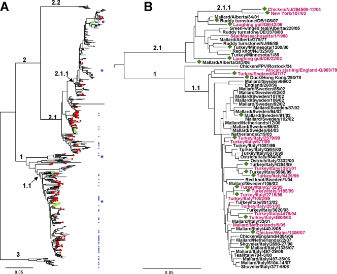
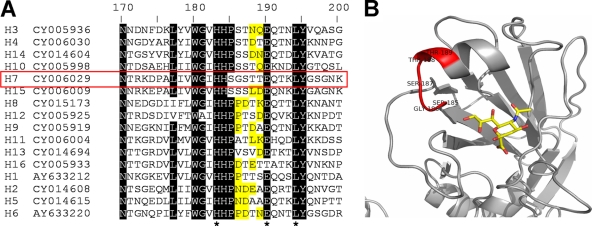
Influenza viruses of gallinaceous poultry and wild aquatic birds usually have distinguishable receptor-binding properties. Here we used a panel of synthetic sialylglycopolymers and solid-phase receptor-binding assays to characterize receptor-binding profiles of about 70 H7 influenza viruses isolated from aquatic birds, land-based poultry, and horses in Eurasia and America. Unlike typical duck influenza viruses with non-H7 hemagglutinin (HA), all avian H7 influenza viruses, irrespective of the host species, displayed a poultry-virus-like binding specificity, i.e., preferential binding to sulfated oligosaccharides Neu5Acα2-3Galβ1-4(6-O-HSO(3))GlcNAc and Neu5Acα2-3Galβ1-4(Fucα1-3)(6-O-HSO(3))GlcNAc. This phenotype correlated with the unique amino acid sequence of the amino acid 185 to 189 loop of H7 HA and seemed to be dependent on ionic interactions between the sulfate group of the receptor and Lys193 and on the lack of sterical clashes between the fucose residue and Gln222. Many North American and Eurasian H7 influenza viruses displayed weak but detectable binding to the human-type receptor moiety Neu5Acα2-6Galβ1-4GlcNAc, highlighting the potential of H7 influenza viruses for avian-to-human transmission. Equine H7 influenza viruses differed from other viruses by preferential binding to the N-glycolyl form of sialic acid. Our data suggest that the receptor-binding site of contemporary H7 influenza viruses in aquatic and terrestrial birds was formed after the introduction of their common precursor from ducks to a new host, presumably, gallinaceous poultry. The uniformity of the receptor-binding profile of H7 influenza viruses in various wild and domestic birds indicates that there is no strong receptor-mediated host range restriction in birds on viruses with this HA subtype. This notion agrees with repeated interspecies transmission of H7 influenza viruses from aquatic birds to poultry.


Figures

Fig 1
Fig 1
Evolutionary relationships of H7 HAs. Phylogenetic trees for the amino acid sequences of the HA1 protein were inferred by the neighbor-joining method using MEGA software version 5 (61). The scale bars represent 0.05 units of amino acid substitutions per site. (A) The tree is based on 665 sequences available from GenBank and 19 sequences determined in this study. Numbered branches include viruses from the Eastern Hemisphere (clade 1) and the Western Hemisphere (clade 2); viruses from North America (clade 2.1) and South America (clade 2.2); viruses isolated in Europe and Asia after 1970 (clade 1.1); North American poultry influenza viruses with an eight-amino-acid deletion in the receptor-binding site (clade 2.1.1); and H7 equine influenza viruses (clade 3). Red dots mark viruses isolated from wild and domestic aquatic birds (mainly ducks). Green dots mark viruses isolated from humans. Blue dots depict viruses tested for receptor binding properties in this study; blue stars show four viruses characterized previously (20). (B) Tree for the viruses tested in this study and in reference . The strain names are colored in accord with the viral receptor-binding specificity: black, typical poultry-virus-like phenotype (Fig. 2B); purple, atypical poultry-virus-like phenotype (Fig. 2C). Green diamonds depict viruses that bind to 6′SLN. The numbering of the clades is the same as in panel A.
Fig 2
Fig 2
Examples of receptor-binding profiles of avian influenza viruses. Association constants of viral complexes with nonlabeled Neu5Ac2-3Gal-containing sialylglycopolymers were determined using a binding inhibition assay. Colors depict the sialyoligosaccharide moiety of the SGP. (A) Duck influenza viruses with non-H7 HA (receptor-binding phenotype d). (B) Viruses representing typical binding phenotype (P) of H7 influenza viruses with a Kass[Su-SLex]/Kass[3′SLN] ratio equal to or higher than 10 (see Table S1 in the supplemental material). (C) H7 influenza viruses with atypical binding phenotypes (p) (Kass[Su-SLex]/Kass[3′SLN] ratio between 1 and 8).
Fig 3
Fig 3
Examples of receptor-binding profiles of equine influenza viruses with H7 and H3 HA. Association constants of viral complexes with nonlabeled sialylglycopolymers were determined using a binding inhibition assay.
Fig 4
Fig 4
Examples of virus binding to Neu5Acα2-3Gal- and Neu5Acα2-6Gal-containing biotinylated SGPs in a direct binding assay. The viruses displayed comparable levels of binding to 3′SLN-containing SGPs (open circles); two of them (panels B and C) also bound to 6′SLN-containing SGPs (closed circles).
Fig 5
Fig 5
Structure of the 185 to 189 peptide loop of H7 HA. (A) Partial amino acid sequences of the 16 HA subtypes. Amino acids in the region from 185 to 189 with bulky side chains are highlighted in yellow. H7 HA is indicated with a red box. GenBank accession numbers for the sequences are shown next to the subtype. The top line shows H3 numbering. Stars in the bottom line depict amino acid residues that interact with sialic acid in the HA-receptor complex. The sequences are listed according to their homology; amino acids conserved among at least 14 of 16 sequences are shaded. (B) Location of the 185 to 189 loop (colored in red) on the model of H7 HA complex with pentasaccharide LSTa (52). Only the sialic acid moiety of LSTa is shown (stick model).
Fig 6
Fig 6
Molecular model of H7 HA complexed with sialyoligosaccharide Su-SLex. The model represents the crystal structure of the H7 HA complex with LSTa (52), in which bound sialyloligosaccharide was modified to generate Su-SLex by the use of Pymol. The HA is shown as a gray molecular surface. Lys193 and Gln222 are highlighted and labeled. The fucose residue of Su-SLex is depicted by purple dots; the sulfo group is shown in orange-red. Panels A and B show different views of the same model and illustrate formation of and ionic bonds between the sulfo group and Lys193 and the lack of sterical conflicts between fucose and Gln222.

Similar articles

Cited by

References

    1. Baigent SJ, McCauley JW. 2003. Influenza type A in humans, mammals and birds: determinants of virus virulence, host-range and interspecies transmission. Bioessays 25:657–671 - PubMed
    1. Banks J, et al. 2001. Changes in the haemagglutinin and the neuraminidase genes prior to the emergence of highly pathogenic H7N1 avian influenza viruses in Italy. Arch. Virol. 146:963–973 - PubMed
    1. Bao Y, et al. 2008. The influenza virus resource at the National Center for Biotechnology Information. J. Virol. 82:596–601 - PMC - PubMed
    1. Belser JA, et al. 2008. Contemporary North American influenza H7 viruses possess human receptor specificity: implications for virus transmissibility. Proc. Natl. Acad. Sci. U. S. A. 105:7558–7563 - PMC - PubMed
    1. Belser JA, Bridges CB, Katz JM, Tumpey TM. 2009. Past, present, and possible future human infection with influenza virus A subtype H7. Emerg. Infect. Dis. 15:859–865 - PMC - PubMed

Publication types

Substances

LinkOut - more resources