. 2012 May;86(9):4833-43.
doi: 10.1128/JVI.05507-11. Epub 2012 Feb 15.

Spike protein VP8* of human rotavirus recognizes histo-blood group antigens in a type-specific manner

Affiliations

Spike protein VP8* of human rotavirus recognizes histo-blood group antigens in a type-specific manner

Pengwei Huang et al. J Virol. 2012 May.

Abstract

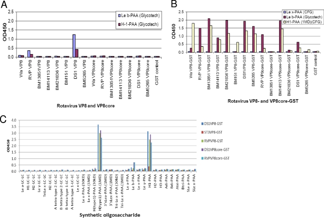
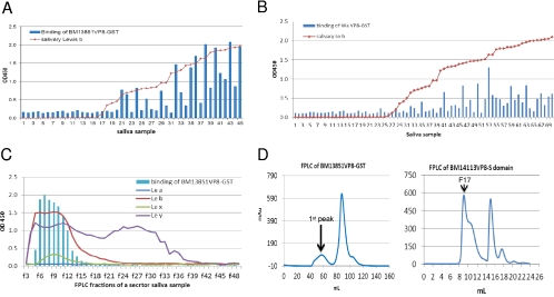
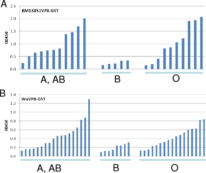
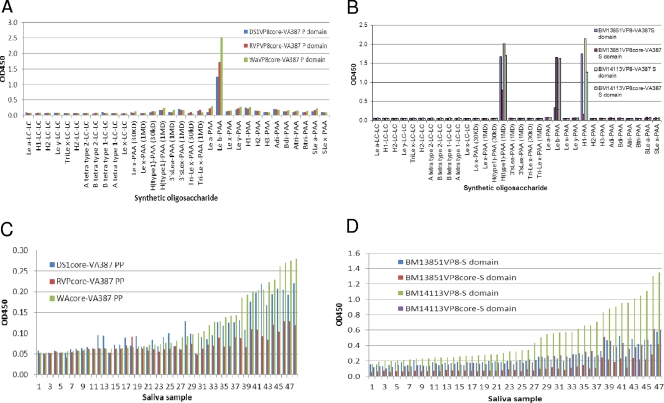
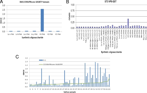
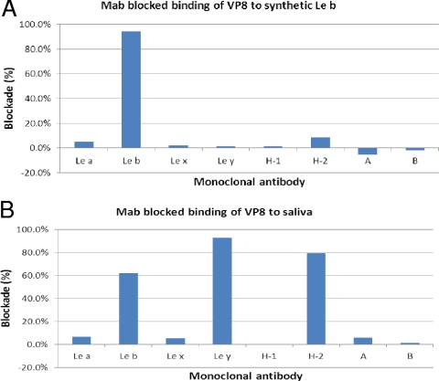
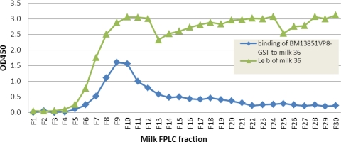
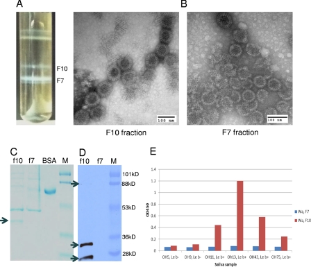
Rotaviruses (RVs), an important cause of severe diarrhea in children, have been found to recognize sialic acid as receptors for host cell attachment. While a few animal RVs (of P[1], P[2], P[3], and P[7]) are sialidase sensitive, human RVs and the majority of animal RVs are sialidase insensitive. In this study, we demonstrated that the surface spike protein VP8* of the major P genotypes of human RVs interacts with the secretor histo-blood group antigens (HBGAs). Strains of the P[4] and P[8] genotypes shared reactivity with the common antigens of Lewis b (Le(b)) and H type 1, while strains of the P[6] genotype bound the H type 1 antigen only. The bindings between recombinant VP8* and human saliva, milk, or synthetic HBGA oligosaccharides were demonstrated, which was confirmed by blockade of the bindings by monoclonal antibodies (MAbs) specific to Le(b) and/or H type 1. In addition, specific binding activities were observed when triple-layered particles of a P[8] (Wa) RV were tested. Our results suggest that the spike protein VP8* of RVs is involved in the recognition of human HBGAs that may function as ligands or receptors for RV attachment to host cells.


Figures

Fig 1
Fig 1
Binding of recombinant RV VP8* to synthetic oligosaccharides. Recombinant proteins with the full-length or the core region of the RV VP8* from seven P[8] or P[4] human RVs were tested for binding to oligosaccharides with (A) or without (B) removal of the GST tag. Except for BM13851 VP8* core-GST to H type 1-PAA and BM13851 VP8* core-GST, BM151 VP8*-GST, and BM5265 VP8* core-GST to Leb-PAA, all VP8*- and VP8* core-GST proteins showed significant binding to Leb and H type 1 antigens compared with binding to Lex-PAA (means + 2 standard deviations [SD]). (C) One of each of the P[4] (DS1) and P[8] (RVP) RVs was tested with extended oligosaccharides representing variable HBGAs from two different sources (GlycoTech Corp. and the CFG) to confirm the binding specificities to the Leb and H type 1 antigens.
Fig 2
Fig 2
Binding of recombinant RV VP8* to saliva. (A and B) VP8*-GST fusion proteins from 2 P[8] (BM13851, Wa) human RVs were tested for binding to a panel of saliva samples. The results for binding of VP8* to individual saliva samples were plotted according to a sorting of the Leb signals of individual saliva samples. Saliva samples were boiled before being used in the assays to remove antibodies fractionation by may interfere with the binding results. A correlation between the salivary Leb and VP8* binding levels for both strains was observed. (C) Binding of an RV VP8* (BM13851) to a secretor saliva sample following the fast-performance liquid chromatography (FPLC). The signals of Lea, Leb, Lex, and Ley of individual fractions were determined by a MAb-based EIA. The VP8*-GST fusion protein bound only to the high-molecular-weight fractions containing the Leb antigen. The chart was made by sorting of binding signals that were presented in line with markers instead of bars indicating correlation with the salivary Lewis antigens. (D) Gel filtration profiles of RV VP8*-GST (Superdex 200 16/60 GL) and VP8*-VA387 S domain fusion proteins (Superdex 200 10/300 GL). The molecular masses of the 1st peak of BM13851 VP8*-GST and fraction 17 (F17) of the BM14113 VP8* S domain were >440 kDa and >800 kDa, respectively.
Fig 3
Fig 3
Characterization of saliva binding signals of RV VP8* based on ABO typing of saliva. The signals for binding of VP8*-GST of BM13851 (A) and Wa (B) to saliva from type A/AB, B, and O secretors (Leb positive) were compared. The low-level binding of VP8* to type B secretor saliva may due to a steric interference from B antigen. The individuals in each of the three groups were sorted from low to high binding strength (OD).
Fig 4
Fig 4
Binding of RV VP8* presented by the NoV VA387 P particle (PP, P domain) and the S complex to synthetic oligosaccharides and saliva. (A) VP8* presented by the NoV P particle bound to the Leb oligosaccharides for both P[8] (Wa and DS1) and P[4] (RVP) RVs. (B) VP8* presented by the NoV S domain bound to the H type 1 in addition to the Leb antigens. (C and D) The bindings of VP8* to a panel of Leb-positive saliva samples were correlated with each other among the VP8* proteins presented by the NoV P particle (C) or the S domain (D).
Fig 5
Fig 5
Binding of VP8* from two P[6] RVs to synthetic oligosaccharides. The ST3 VP8*-GST fusion protein and the BM11596 VP8* core protein presented by the VA387 P domain (P particle, PP) were tested by the oligosaccharide binding assays described in Materials and Methods. Both BM11596 (A) and ST3 (B) recognized PAA-conjugated H type 1 only. (C) A panel of Lewis b-negative saliva samples was tested for binding to BM11596 VP8*. The data for individual saliva samples were sorted by their binding activities with the VP8* protein.
Fig 6
Fig 6
Blocking of binding of RV VP8* to synthetic oligosaccharide (A) and saliva (B) by monoclonal antibodies (MAbs) specific for HBGAs. Microtiter plates were coated with Leb oligosaccharide (A) or Leb-positive saliva (B) to capture RV VP8*-GST fusion protein (∼10 μg/ml) with or without preincubation with MAbs (at a dilution of 1:20) against Lea, Leb, Lex, Ley, A, B, H type 1, and H type 2 antigens. VP8*-specific blocking activity was determined by the reduction (%) of the optical density values in wells with MAbs compared with those in wells without MAbs.
Fig 7
Fig 7
Binding of RV-VP8* to milk Leb antigen in size-exclusive FPLC fractions. Boiled FPLC fractions of one secretor's milk sample (milk 36) were used to coat microtiter plates at a dilution of 1:15 in 1× PBS. The bound BM13851 VP8*-GST protein was detected by rotavirus hyperimmune sera. Only the high-molecular-weight fractions containing Leb antigen showed binding. The distribution of milk Leb in the FPLC fraction was previously measured in a separate experiment.
Fig 8
Fig 8
Purification of Wa TLP and its binding to saliva samples from Leb-positive individuals. Wa TLP and DLP bands were observed in a CsCl gradient purification (A). DLPs (F7; density, 1.39 g/cm3 in CsCl) and TLPs (F10; density, 1.36 g/cm3 in CsCl) were confirmed by electron microscopy (EM) examination (B) and Western blot analysis (D). Typical TLPs and DLPs were observed in each of the two fractions by EM in negatively stained grids (B). SDS-PAGE followed by Coomassie blue staining revealed an extra protein band (38 kDa) (arrow) in F10 but not in F7, which is predicted to be the VP7 protein on the outer layer of TLPs, missing on DLPs. The amounts of viral loading of F10 and F7 in the gels were adjusted to equality on the basis of the amounts of VP6 in each of the two fractions (C). The presence of the other major outer layer surface protein VP4 was confirmed by detection of VP4 (top arrow), the protease-processed VP8* protein (middle arrow), and a truncated VP8* protein (bottom arrow) by Western blot analysis using a VP8*-specific monoclonal antibody (D). Specific binding of the Wa TLPs, but not the DLPs, to the Leb HBGAs was demonstrated by the saliva-based binding assay using a panel of Leb-positive and -negative saliva samples (E). Equal volumes (10 μl) of F7 and F10 were used in the saliva binding assay. A serial dilution analysis by SDS-PAGE showed that the amount of the DLPs was ∼4-fold larger than that of the TLPs based on the intensities of the major structural protein VP6 in the two preps in the gel (data not shown).

References

    1. Ahmed T, et al. 2009. Children with the Le(a+b-) blood group have increased susceptibility to diarrhea caused by enterotoxigenic Escherichia coli expressing colonization factor I group fimbriae. Infect. Immun. 77:2059–2064 - PMC - PubMed
    1. Arias CF, et al. 2002. Molecular biology of rotavirus cell entry. Arch. Med. Res. 33:356–361 - PubMed
    1. Bahl R, et al. 2005. Incidence of severe rotavirus diarrhea in New Delhi, India, and G and P types of the infecting rotavirus strains. J. Infect. Dis. 192(Suppl. 1):S114–S119 - PubMed
    1. Beards G, Graham C. 1995. Temporal distribution of rotavirus G-serotypes in the West Midlands region of the United Kingdom, 1983–1994. J. Diarrhoeal Dis. Res. 13:235–237 - PubMed
    1. Beards GM, Desselberger U, Flewett TH. 1989. Temporal and geographical distributions of human rotavirus serotypes, 1983 to 1988. J. Clin. Microbiol. 27:2827–2833 - PMC - PubMed

Publication types

MeSH terms

LinkOut - more resources