. 2012 May;86(9):5039-54.
doi: 10.1128/JVI.07162-11. Epub 2012 Feb 15.

Biological characterization and next-generation genome sequencing of the unclassified Cotia virus SPAn232 (Poxviridae)

Affiliations

Biological characterization and next-generation genome sequencing of the unclassified Cotia virus SPAn232 (Poxviridae)

Priscila P Afonso et al. J Virol. 2012 May.

Abstract

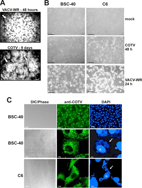
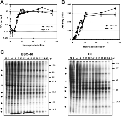
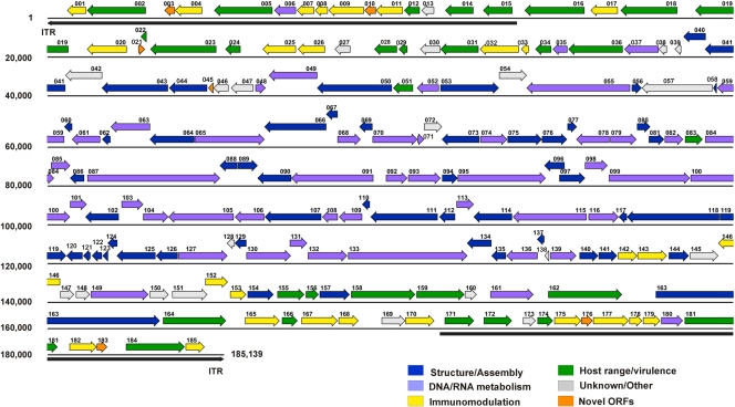
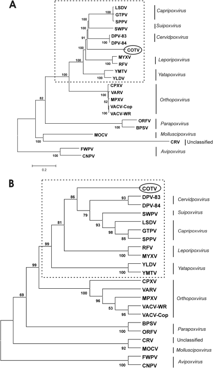
Cotia virus (COTV) SPAn232 was isolated in 1961 from sentinel mice at Cotia field station, São Paulo, Brazil. Attempts to classify COTV within a recognized genus of the Poxviridae have generated contradictory findings. Studies by different researchers suggested some similarity to myxoma virus and swinepox virus, whereas another investigation characterized COTV SPAn232 as a vaccinia virus strain. Because of the lack of consensus, we have conducted an independent biological and molecular characterization of COTV. Virus growth curves reached maximum yields at approximately 24 to 48 h and were accompanied by virus DNA replication and a characteristic early/late pattern of viral protein synthesis. Interestingly, COTV did not induce detectable cytopathic effects in BSC-40 cells until 4 days postinfection and generated viral plaques only after 8 days. We determined the complete genomic sequence of COTV by using a combination of the next-generation DNA sequencing technologies 454 and Illumina. A unique contiguous sequence of 185,139 bp containing 185 genes, including the 90 genes conserved in all chordopoxviruses, was obtained. COTV has an interesting panel of open reading frames (ORFs) related to the evasion of host defense, including two novel genes encoding C-C chemokine-like proteins, each present in duplicate copies. Phylogenetic analysis revealed the highest amino acid identity scores with Cervidpoxvirus, Capripoxvirus, Suipoxvirus, Leporipoxvirus, and Yatapoxvirus. However, COTV grouped as an independent branch within this clade, which clearly excluded its classification as an Orthopoxvirus. Therefore, our data suggest that COTV could represent a new poxvirus genus.


Figures

Fig 1
Fig 1
Electron microscopy analysis of COTV-infected cells. C6 (A, B, and D) and BSC-40 (C) cells were infected with COTV at an MOI of 1. The monolayers were fixed and processed for transmission electron microscopy at 24 h (A) or 96 h (B to D) postinfection. Representative fields are shown. (A) Mature viruses (MV) and immature spherical particles (IV) are visualized in the cytoplasm outside areas devoid of cellular organelles resembling Cotia bodies (asterisks). Extracellular viruses (EV) are found associated with the cell membrane. (Inset) High-magnification image of viroplasm (Vi) surrounded by membrane crescents (Cr) and IV. (B) High-magnification image of MV. (C) MV are enveloped by additional membranes derived from the trans-Golgi complex, generating wrapped virions (WV). The Golgi apparatus (Go) is shown. (D) High-magnification image of EVs associated with the cell membrane. Bars, 200 nm.
Fig 2
Fig 2
Virus plaque phenotype and CPE progression in COTV-infected cells. (A) Monolayers of BSC-40 cells were infected with 300 PFU of VACV-WR or COTV for 2 or 9 days, respectively, at which time the cells were stained with 0.1% crystal violet. Arrows indicate viral plaques. Bars, 100 μm. (B) BSC-40 and C6 cells were either mock infected or infected with COTV or VACV-WR at an MOI of 1, and CPE was visualized at 24 (VACV-WR) or 48 (COTV) h postinfection. Bars, 100 μm. (C) BSC-40 and C6 cells were infected with COTV at an MOI of 1 for 48 h and were processed for immunofluorescence assays using an antiserum against COTV structural proteins (anti-COTV). DNA was stained with DAPI. N, nucleus. Arrows point to virus factories in the cell cytoplasm. Representative fields are shown. Bars, 50 μm (top) and 5 μm (center and bottom).
Fig 3
Fig 3
Time course analysis of progeny production, DNA replication, and protein synthesis in COTV-infected cells. Semiconfluent BSC-40 and C6 monolayers were infected with COTV at an MOI of 1, and at the indicated times postinfection, cells were either harvested for virus titration by plaque assay (A), processed for detection of viral DNA by slot blot hybridization (B), or pulse-labeled with [35S]methionine followed by 12% SDS-PAGE analysis (C). (A) Values represent the means for three assays titrated in duplicate. (B) The autoradiograms obtained for BSC-40 and C6 cells were scanned, and densitometry analysis was performed. The numbers (arbitrary units) express the mean values for nine autoradiograms in which samples were applied in triplicate. (C) Representative autoradiograms are shown. Filled circles indicate viral late proteins; arrowheads indicate viral early proteins; asterisks indicate a host protein. M, mock-infected cells. Molecular size markers (in kilodaltons) are given on the right.
Fig 4
Fig 4
Analysis of COTV cross-reactivity and virus neutralization. (A) The indicated cells were infected (INF) with COTV (BSC-40), VACV (BSC-40), MYXV (RK-13), or SWPV (PK-15) at an MOI of 5 and were harvested after reaching intense CPE (for COTV, 4 days; for VACV, 24 h; for MYXV, 48 h; for SWPV, 3 days). Samples were analyzed by Western blotting using an antiserum against COTV (left), VACV (center), or MYXV (right). (B) A total of 10,000 PFU of COTV, VACV, MYXV, or SWPV was incubated with the indicated dilutions of anti-COTV antiserum or the preimmune serum (PI) for 1 h at 37°C. The mixtures were then placed on monolayers of BSC-40 (COTV and VACV), RK-13 (MYXV), or PK-15 (SWPV) cells for 9 days (COTV) or for 48, 52, or 96 h postinfection (VACV, MYXV, or SWPV, respectively), after which virus-induced CPE was visualized by crystal violet staining.
Fig 5
Fig 5
ORF map of the COTV genome. The annotated ORFs are represented by arrows color coded according to their functional categories. The arrows representing ORFs point left or right, indicating the direction of transcription. The inverted terminal repeat (ITR) regions are indicated by long black arrows (shown below the sequence) at the ends of the genome.
Fig 6
Fig 6
Amino acid alignment of COTV C-C motif chemokine-like proteins (COTV003 and COTV010) with cellular homologs. The chemokine superfamily domain detected by Pfam, Smart, and InterPro is shaded. The four conserved cysteine residues involved in the formation of two disulfide bonds are boxed. Amino acid positions are indicated on the right. GenBank accession numbers are as follows: Mustela putorius furo (ferret) C-C motif chemokine ligand 13, ACJ54430.1; Mus musculus (mouse) small inducible cytokine A2 precursor, AAF15379.1; Pan troglodytes (chimpanzee) C-C motif chemokine 4 isoform 3, XP_001173914; Homo sapiens (human) Act-2 cytokine, AAB00790; Oryctolagus cuniculus (rabbit) C-C motif chemokine ligand 3-like, XP_002719292.
Fig 7
Fig 7
Phylogenetic inference of COTV. The concatenated data set was obtained by combining the individual alignments of the predicted amino acid sequences for 90 genes conserved in 22 chordopoxviruses (ORFs in boldface in Table 2). The combined alignment was used to construct the neighbor-joining tree, opting for the JTT model of substitution, with 2,500 bootstrap replicates, using MEGA 4 (A), and the maximum-likelihood tree, opting for WAG correction for multiple substitutions, 10,000 quartet puzzling steps, and the gamma heterogeneity model (B). The bar in panel A represents relative genetic distance. The poxvirus genera and subfamilies are given on the right. Dashed boxes enclose the poxvirus CSYLC clade, which includes Capripoxvirus, Suipoxvirus, Yatapoxvirus, Leporipoxvirus, and Cervidpoxvirus.

References

    1. Afonso CL, et al. 2005. Genome of deerpox virus. J. Virol. 79:966–977 - PMC - PubMed
    1. Afonso CL, et al. 2006. Genome of crocodilepox virus. J. Virol. 80:4978–4991 - PMC - PubMed
    1. Afonso CL, et al. 2000. The genome of fowlpox virus. J. Virol. 74:3815–3831 - PMC - PubMed
    1. Afonso CL, et al. 2002. The genome of swinepox virus. J. Virol. 76:783–790 - PMC - PubMed
    1. Afonso CL, et al. 2002. The genome of camelpox virus. Virology 295:1–9 - PubMed

Publication types

MeSH terms

Associated data

LinkOut - more resources