. 2012 Apr;86(8):4058-64.
doi: 10.1128/JVI.00012-12. Epub 2012 Feb 15.

The UL36 tegument protein of herpes simplex virus 1 has a composite binding site at the capsid vertices

Affiliations

The UL36 tegument protein of herpes simplex virus 1 has a composite binding site at the capsid vertices

Giovanni Cardone et al. J Virol. 2012 Apr.

Abstract

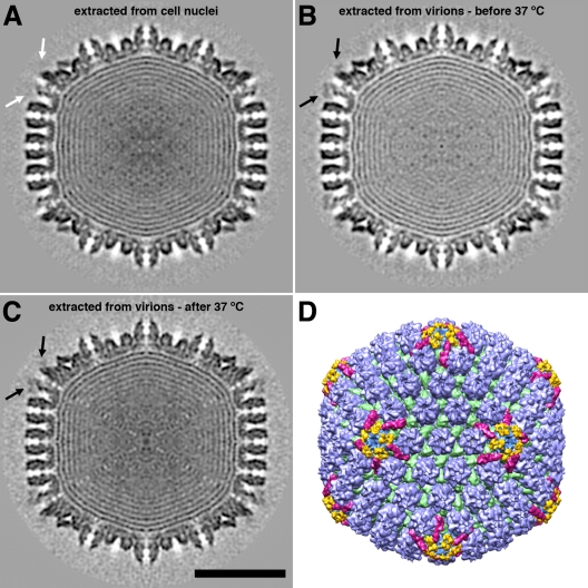
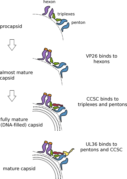
Herpesviruses have an icosahedral nucleocapsid surrounded by an amorphous tegument and a lipoprotein envelope. The tegument comprises at least 20 proteins destined for delivery into the host cell. As the tegument does not have a regular structure, the question arises of how its proteins are recruited. The herpes simplex virus 1 (HSV-1) tegument is known to contact the capsid at its vertices, and two proteins, UL36 and UL37, have been identified as candidates for this interaction. We show that the interaction is mediated exclusively by UL36. HSV-1 nucleocapsids extracted from virions shed their UL37 upon incubation at 37°C. Cryo-electron microscopy (cryo-EM) analysis of capsids with and without UL37 reveals the same penton-capping density in both cases. As no other tegument proteins are retained in significant amounts, it follows that this density feature (∼100 kDa) represents the ordered portion of UL36 (336 kDa). It binds between neighboring UL19 protrusions and to an adjacent UL17 molecule. These observations support the hypothesis that UL36 plays a major role in the tegumentation of the virion, providing a flexible scaffold to which other tegument proteins, including UL37, bind. They also indicate how sequential conformational changes in the maturing nucleocapsid control the ordered binding, first of UL25/UL17 and then of UL36.


Figures

Fig 1
Fig 1
Cryo-electron micrographs of HSV-1 T36 capsids isolated at 4°C (A) and at 37°C (B). Bar = 100 nm.
Fig 2
Fig 2
SDS-polyacrylamide gel electrophoresis comparing the protein compositions of purified HSV-1 virus (left lane) with T36 capsids prepared at 4°C (right) and at 37°C (middle). The molecular masses of the proteins in the bands labeled to the right of the gel are as follows: 336 kDa for UL36, 149 kDa for UL19, 121 kDa for UL37, 75 kDa for UL17, 63 kDa for UL25, and 50 kDa for UL38. UL36 is the virion protein with the highest mass. The UL37 band was identified by Western blotting and mass spectrometry (W. W. Newcomb, L. M. Jones, A. Dee, and J. C. Brown, unpublished results).
Fig 3
Fig 3
Three-dimensional reconstructions of DNA-containing HSV-1 capsids. (A to C) Central sections through capsids viewed along a 2-fold axis of icosahedral symmetry. (A) Nuclear C-capsid; (B) T36 capsid isolated at 4°C; (C) T36 capsid isolated at 37°C. The black arrows in panels B and C point to additional density features overlying pentons. There is no such density feature in panel A (white arrow). Bar = 50 nm. (D) Color-coded surface rendering of the reconstruction shown in panel C. The surface features are shown in color as follows: capsomer protrusions are blue, triplexes are green, CCSCs are magenta, and the additional penton-associated density features (part of UL36) found on the T36 capsid are yellow.
Fig 4
Fig 4
Higher-magnification view of the region surrounding a vertex on the reconstruction of the T36 capsid isolated at 4°C, both before (A) and after (B) incubation at 37°C. The surface features were color coded as in Fig. 3. Bar = 10 nm.
Fig 5
Fig 5
T36 capsids were analyzed by content of UL36. (A and C) T36 capsid subset with the highest level of occupancy; (B and D) T36 capsid subset with the lowest level of occupancy. The UL36-related density is indicated by black arrows in panel A and gray arrows in panel B. The surface features in panels C and D are color coded as in Fig. 3. Bar = 20 nm.
Fig 6
Fig 6
Cartoon summarizing regulation of the sequential addition of proteins to the HSV-1 capsid. In the first step, as the procapsid undergoes initial maturation, its shape alters from round to polyhedral, and the hexon protrusions close up and become 6-fold symmetric, creating binding sites for VP26. Late in DNA packaging, another less extreme transition takes place that affects the peripentonal triplexes and/or the penton so as to enhance the affinity of this composite binding site for the CCSC, whose binding promotes nuclear exit. Subsequently, (11, 20), UL36 binds to another composite site involving both the CCSC and UL19 penton tips. In this way, UL25/UL17 plays a functional role in the cytoplasm similar to that which it plays in the nucleus, allowing only DNA-filled capsids to progress efficiently along the exit pathway.

References

    1. Baines JD. 2007. Envelopment of herpes simplex virus nucleocapsids at the inner membrane, p 144–150 In Arvin A, et al. (ed), Human herpesviruses: biology, therapy, and immunoprophylaxis. Cambridge University Press, Cambridge, United Kingdom
    1. Booy FP, et al. 1994. Finding a needle in a haystack: detection of a small protein (the 12-kDa VP26) in a large complex (the 200-MDa capsid of herpes simplex virus). Proc. Natl. Acad. Sci. U. S. A. 91:5652–5656 - PMC - PubMed
    1. Britt B. 2007. CMV maturation and egress, p 311–323 In Arvin A, et al. (ed), Human herpesviruses: biology, therapy, and immunoprophylaxis. Cambridge University Press, Cambridge, United Kingdom - PubMed
    1. Bubeck D, et al. 2005. Structure of the poliovirus 135S cell entry intermediate at 10-angstrom resolution reveals the location of an externalized polypeptide that binds to membranes. J. Virol. 79:7745–7755 - PMC - PubMed
    1. Cardone G, Heymann JB, Cheng N, Trus BL, Steven AC. 2012. Procapsid assembly, maturation, nuclear exit: dynamic steps in the production of infectious herpesvirions, p 423–440 In Rossmann MG, Rao VB. (ed), Viral molecular machines. Springer, New York, NY - PMC - PubMed

Publication types

MeSH terms