. 2012 Mar 15;188(6):2794-804.
doi: 10.4049/jimmunol.1102068. Epub 2012 Feb 15.

Human cytomegalovirus UL40 signal peptide regulates cell surface expression of the NK cell ligands HLA-E and gpUL18

Affiliations

Human cytomegalovirus UL40 signal peptide regulates cell surface expression of the NK cell ligands HLA-E and gpUL18

Virginie Prod'homme et al. J Immunol. .

Abstract

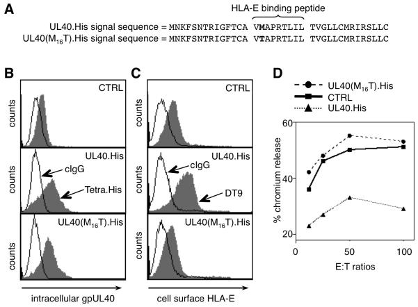
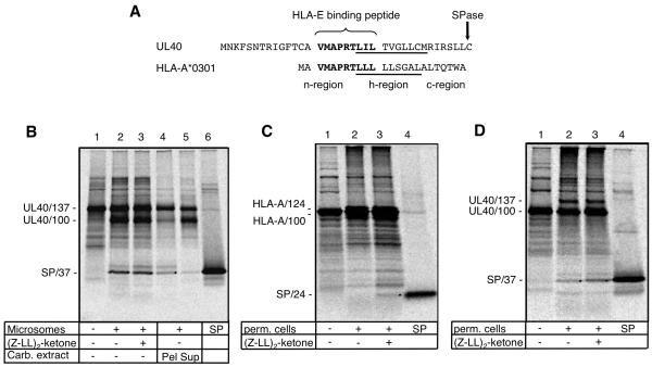
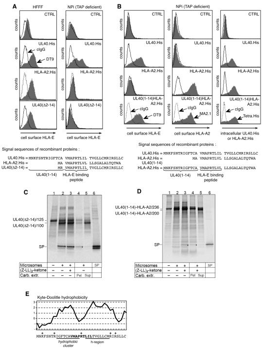
Human CMV (HCMV)-encoded NK cell-evasion functions include an MHC class I homolog (UL18) with high affinity for the leukocyte inhibitory receptor-1 (CD85j, ILT2, or LILRB1) and a signal peptide (SP(UL40)) that acts by upregulating cell surface expression of HLA-E. Detailed characterization of SP(UL40) revealed that the N-terminal 14 aa residues bestowed TAP-independent upregulation of HLA-E, whereas C region sequences delayed processing of SP(UL40) by a signal peptide peptidase-type intramembrane protease. Most significantly, the consensus HLA-E-binding epitope within SP(UL40) was shown to promote cell surface expression of both HLA-E and gpUL18. UL40 was found to possess two transcription start sites, with utilization of the downstream site resulting in translation being initiated within the HLA-E-binding epitope (P2). Remarkably, this truncated SP(UL40) was functional and retained the capacity to upregulate gpUL18 but not HLA-E. Thus, our findings identify an elegant mechanism by which an HCMV signal peptide differentially regulates two distinct NK cell-evasion pathways. Moreover, we describe a natural SP(UL40) mutant that provides a clear example of an HCMV clinical virus with a defect in an NK cell-evasion function and exemplifies issues that confront the virus when adapting to immunogenetic diversity in the host.


Figures

Figure 1
Figure 1. Consensus VMAPRTLIL sequence implicated in UL40 function
RAd-UL40.His has a wild type HLA-E-binding epitope (VMAPRTLIL), whereas RAd-UL40(M16T).His has a mutated form (VTAPRTLIL). RAd-CTRL is the same vector without a transgene. (A) The signal peptide present in each construct showing the M/T mutation in bold. (B) Intracellular expression of gpUL40 and (C) cell surface expression of HLA-E*0101 in HFFF infected with RAd-CTRL, RAd-UL40.His and RAd-UL40(M16T).His measured using antibodies to the C-terminal epitope tag and to HLA-E/-C (DT9), respectively. (D) An NK cell cytolysis assay was performed using NKL cells incubated with HFFF cells infected with the RAds as indicated. Results are representative of three independent experiments.
Figure 2
Figure 2. Modulation of HLA-E expression by natural UL40 sequence variants
Cell surface expression of HLA-E as measured by the mAb DT9 following (A) infection of HFFF with HCMV strains AD169, Toledo and 3157 (72 h post infection). Downregulation of MHC-I (mAb W6/32) was used as a marker of HCMV infection. (B) Infection of HFFF with RAds expressing UL40 from different strains. The sequences of SPUL40 from the different virus strains are shown.
Figure 3
Figure 3. Processing of HLA-A*0301 and UL40 signal peptides
(A) Alignment of the UL40 and HLA-A*0301 SPs illustrating the SPase cleavage sites plus the n, h (hydrophobic; underlined) and c sequences in HLA-A*0301 (B) In vitro translation of mRNA coding for the signal peptide plus 100 aa of UL40 (UL40/137; lane 1) in the presence of ER-derived microsomes (lanes 2-5) and (Z-LL)2-ketone (lane 3). One aliquot of microsomes was extracted with sodium carbonate and separated into pellet (Pel, lane 4) and supernatant (Sup, lane 5). Dots indicate the SPs; lane 6 shows in vitro-translated UL40 SP. (C) and (D) In vitro translation of mRNA coding for the signal peptide plus 100 aa of HLA-A*0301 (HLA-A/124; lane 1) or UL40 (UL40/137; lane 1) in the presence of selectively permeabilized 721.221 cells (lanes 2-3) and (Z-LL)2-ketone (lane 3). Dots indicate the SPs; lane 4 shows in vitro-translated reference SP.
Figure 4
Figure 4. The SPUL40 ‘n-region’ mediates TAP-independent HLA-E cell surface expression
(A) HFFF (TAP+) or NPi (TAP) cells infected with RAd-CTRL, RAd-UL40.His, RAd-HLA-A2.His or RAd-UL40(Δ2-14). Cell surface expression of HLA-E was assessed using the mAb DT9. (B) NPi cells were infected with RAd-CTRL, RAd-UL40.His, RAd-HLA-A2.His or RAd-UL40(1-14)HLA-A2.His and analyzed for cell surface expression of HLA-E, HLA-A2 or intracellular expression of His-tagged proteins using the indicated mAbs. The signal peptide structure of each construct is shown. Results are representative of three independent experiments. In vitro translation of mRNA coding for (C) the signal peptide plus 100 aa of UL40(Δ2-14) (UL40(Δ2-14)/125; lane 1) or (D) the signal peptide plus 200 aa of UL40(1-14)HLA-A2 (UL40(1-14)HLA-A2/236, lane 1) in the presence of ER-derived microsomes (lanes 2-5) and (Z-LL)2-ketone as indicated. One aliquot of microsomes was extracted with sodium carbonate and separated into pellet (Pel, lane 4) and supernatant (Sup, lane 5). Dots indicate the SPs; lanes 6 show in vitro-translated SPs. (E) Hydrophobicity plot of the SPUL40 (strain AD169) illustrating in addition to the canonical h-region, which is the prime determinant for ER targeting and insertion of the nascent chain, an additional hydrophobic cluster within the n-region. The HLA-E/gpUL18-binding peptide is indicated in bold; position of positive charged amino acid residues with a +.
Figure 5
Figure 5. UL40 promotes cell surface expression of gpUL18 and HLA-E
(A) Cell surface expression of gpUL18, MHC-I, gB, gH and gN compared on HFFF cells infected with HCMV strain AD169 or AD169ΔUL40 for 72h (30 PFU/cell). (B) Cell surface expression of gpUL18 on cells infected with HCMV strain AD169, AD169ΔUL40, AD169ΔUL18 (30 PFU/cell) for 72h. (C) gpUL18 surface expression on cells co-infected with RAd-UL18 (100 PFU/cell) and either RAd-UL40 or the vector control (RAd-CTRL) (30 PFU/cell) for 72h. (D) HFFF infected with RAd-UL18 at the multiplicities of infection (m.o.i) indicated and RAd-UL40 or RAd-CTRL (30 PFU/cell) for 72h. Results are means ± SEM of duplicate samples (Two-Way ANOVA test with Bonferroni post-tests). (E) Cell surface expression of HLA-E (antibody 3D12) and MHC-I on HFFF infected with strain AD169ΔUL16, AD169ΔUL18 or AD169ΔUL40 (15 PFU/cell) for 96h (F) Western blot of HFFF infected with AD169ΔUL16, AD169ΔUL18 or AD169ΔUL40 (15 PFU/cell) for 96h. (G) gpUL18 detected by western blot in HFFF infected with RAd-UL18 (100 PFU/cell) and either RAd-UL40 or RAd-CTRL (30 PFU/cell) for 72h. Cell extracts were digested with Endo H [E] or PNGase F [P] or no enzyme [Mock; M]. Endo H-resistant gpUL18 glycoform (formula image), Endo H-sensitive gpUL18 glycoform before (formula image) and after (formula image) digestion, PNGase-digested gpUL18 forms (formula image) are indicated. (H) Following cell surface biotinylation and immunoprecipitation, gpUL18, HLA-E, HLA-HC and CD155 cell surface expression were compared on western blot in cells infected with AD169ΔUL16, AD169ΔUL18 or AD169ΔUL40 (15 PFU/cell) for 96h.
Figure 6
Figure 6. SPUL40 signal enhances cell surface expression of gpUL18
Cell surface expression of gpUL18 was monitored by flow cytometry on HFFF cells infected for 72 h with the RAds indicated. RAd-UL18 (100 PFU/cell) plus RAd-CTRL (30 PFU/cell) and RAd-UL18 plus RAd-UL40 (30 PFU/cell) were used as standards in all panels. RAd-UL18 plus a second RAd (30 PFU/cell) encoding: (A) SPUL40 cloned in-frame with RFP, (B) SPHLA-A2 in-frame with RFP, (C) UL40 with residues 24-37 replaced with residues 12-24 from HLA-A2, (D) UL40 with residues 1-14 replaced with first 2 residues from HLA-C, (E) UL40 with HLA-E-binding peptide replaced with an HIV-1 GAG epitope, (F) strain Toledo UL40, (G) UL40 with the M16T mutation, or (H) strain 3157 UL40. Results are representative of three independent experiments.
Figure 7
Figure 7. UL40 transcriptional analysis
(A) Northern blot of a strand-specific UL40 probe hybridized to strain Merlin early (E) and late (L) RNAs. The size range (in nt) of the UL40 mRNAs is indicated on the right. Larger RNAs that are 3′-coterminal with UL40 were also detected: major species at approximately 1400 nt (UL41A) and 4500 nt (UL44), plus less abundant species. (B) Agarose gel of RACE products generated from the 5′- and 3′-ends of strain Merlin E and L RNAs using UL40-specific primers. Approximate sizes of the UL40 products from 3′-RACE (560 bp) and 5′-RACE (420 and 310 bp, each with an artefactual, more slowly migrating band above) are shown on the left. DNA markers (M) are shown on the right (sizes in bp). The larger products at approximately 900 bp in the 5′-L lane represent the 5′-end of an upstream, 3′-coterminal gene (UL41A), which maps to residue 54804 in the Merlin sequence, downstream from a potential TATA box (TATATT). (C) Locations of the 5′- and 3′-ends of UL40 mRNAs aligned with the relevant DNA and amino acid sequences (predicted SPs). Dots indicate the central portion of the UL40 coding region, which is not shown. The major 5′-ends are in bold, underlined type and marked with broad arrows. The upstream and downstream occurrences were identified from 21/25 and 23/27 clones, respectively. The ATG codons that initiate the long and short forms of the UL40 protein are in bold, underlined type; an upstream ATG in another reading frame is also underlined. The major 3′-ends are in bold, underlined type and marked by narrow arrows. Their occurrences, as read from left to right, were identified from 3, 7 and 4 clones (from a total of 18 clones), and are located downstream from the bold, underlined canonical polyadenylation signal (AATAAA).
Figure 8
Figure 8. Postulated sequential topogenesis and processing of the SP UL40
Upon targeting and translocation, the gpUL40 precursor (pre-gpUL40) is cleaved by SPase. The liberated SP (SPUL40) remains anchored in the membrane. Proposed association of the hydrophobic N-terminal segment (yellow box labeled *) with the membrane, and subsequent flipping of the hydrophilic spacer (green) and the c-region (blue) are indicated. The canonical n- and h-region are indicted in yellow (h) and red (n), respectively. In semi-intact cells, SPUL40 is further processed by an SPP-type intramembrane protease. The consensus HLA-E-binding peptide is delivered to the ER in a TAP-independent manner, where it binds and stabilizes both endogenous HLA-E and the HCMV-encoded MHC-I homologue gpUL18.

References

    1. McSharry BP, Burgert HG, Owen DP, Stanton RJ, Prod’homme V, Sester M, Koebernick K, Groh V, Spies T, Cox S, Little AM, Wang EC, Tomasec P, Wilkinson GW. Adenovirus E3/19K promotes evasion of NK cell recognition by intracellular sequestration of the NKG2D ligands major histocompatibility complex class I chain-related proteins A and B. J Virol. 2008;82:4585–4594. - PMC - PubMed
    1. Mocarski ES., Jr. Immunomodulation by cytomegaloviruses: manipulative strategies beyond evasion. Trends Microbiol. 2002;10:332–339. - PubMed
    1. Wilkinson GW, Tomasec P, Stanton RJ, Armstrong M, Prod’homme V, Aicheler R, McSharry BP, Rickards CR, Cochrane D, Llewellyn-Lacey S, Wang EC, Griffin CA, Davison AJ. Modulation of natural killer cells by human cytomegalovirus. J Clin Virol. 2008;41:206–212. - PMC - PubMed
    1. Hansen SG, Powers CJ, Richards R, Ventura AB, Ford JC, Siess D, Axthelm MK, Nelson JA, Jarvis MA, Picker LJ, Fruh K. Evasion of CD8+ T cells is critical for superinfection by cytomegalovirus. Science. 2010;328:102–106. - PMC - PubMed
    1. Yokoyama WM, Kim S, French AR. The dynamic life of natural killer cells. Annu Rev Immunol. 2004;22:405–429. - PubMed

Publication types

MeSH terms

Substances