. 2012 May;53(5):909-917.
doi: 10.1194/jlr.M025403. Epub 2012 Feb 16.

FSH stimulates lipid biosynthesis in chicken adipose tissue by upregulating the expression of its receptor FSHR

Affiliations

FSH stimulates lipid biosynthesis in chicken adipose tissue by upregulating the expression of its receptor FSHR

Huanxian Cui et al. J Lipid Res. 2012 May.

Abstract

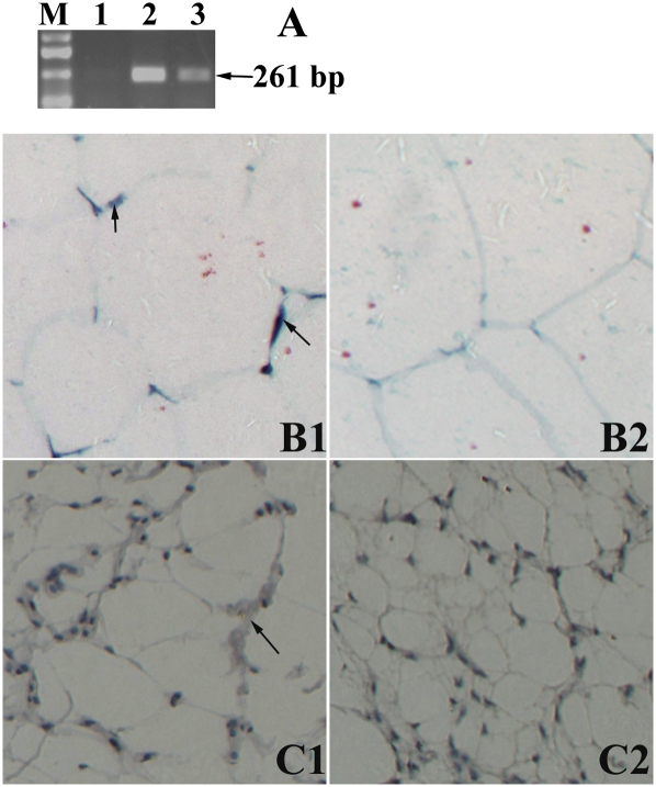
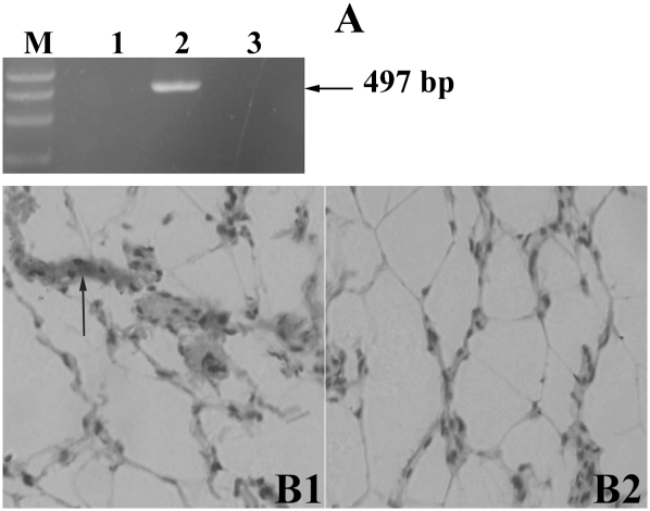
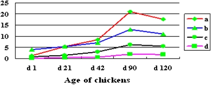
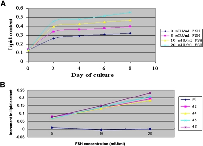
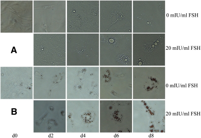
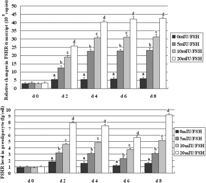
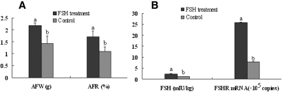
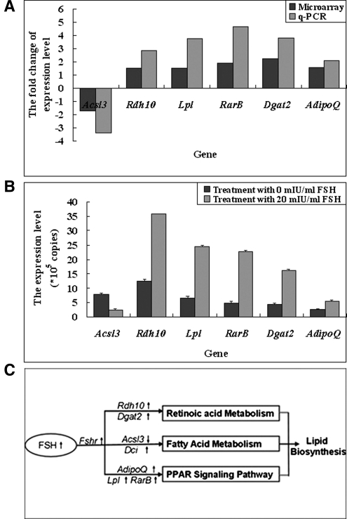
Transcripts and protein for follicle-stimulating hormone receptor (FSHR) were demonstrated in abdominal adipose tissue of female chickens. There was no expression of the Fsh gene, but FSH and FSHR colocalized, suggesting that FSH was receptor bound. Partial correlations indicted that changes in abdominal fat (AF) content were most directly correlated with Fshr mRNA expression, and the latter was directly correlated with tissue FSH content. These relationships were consistent with FSH inducing Fshr mRNA expression and with the finding that FSH influenced the accumulation of AF in chickens, a novel role for the hormone. Chicken preadipocytes responded linearly to doubling concentrations of FSH in Fshr mRNA expression and quantities of FSHR and lipid, without discernable effect on proliferation. Cells exposed to FSH more rapidly acquired adipocyte morphology. Treatment of young chickens with chicken FSH (4 mIU/day, subcutaneous, days 7-13) did not significantly decrease live weight but increased AF weight by 54.61%, AF as a percentage of live weight by 55.45%, and FSHR transcripts in AF by 222.15% (2 h after injection). In cells stimulated by FSH, genes related to lipid metabolism, including Rdh10, Dci, RarB, Lpl, Acsl3, and Dgat2, were expressed differentially, compared with no FSH. Several pathways of retinal and fatty acid metabolism, and peroxisome proliferator-activated receptor (PPAR) signaling changed. In conclusion, FSH stimulates lipid biosynthesis by upregulating Fshr mRNA expression in abdominal adipose tissue of chickens. Several genes involved in fatty acid and retinal metabolism and the PPAR signaling pathway mediate this novel function of FSH.


Figures

Fig. 1.
Fig. 1.
Expression of Fshr mRNA in chicken abdominal adipose tissue. A: Fshr mRNA expression was detected by RT-PCR. Lanes 1–3: negative control, positive control (ovary), abdominal adipose tissue, respectively. M, DL2000 Marker. The expected 261 bp product was verified by electrophoresis in abdominal adipose tissue. B1: Fshr mRNA ISH: positive hybridized signals (arrows) were showed in abdominal adipose tissue, which indicate expression of Fshr mRNA (inverted microscope, 200×). C1: immunocytochemical detection of FSHR protein in abdominal adipose tissue: positive signal appears as brown staining surrounding the fat locule (inverted microscope, 200×). B2 and C2, the negative controls.
Fig. 2.
Fig. 2.
Fsh gene is not expressed but the hormone is detectable in chicken abdominal adipose tissue. A: Fsh mRNA expression was detected by RT-PCR. Lanes 1–3: negative control, positive control (pituitary), and abdominal adipose tissue, respectively. M, DL2000 marker. No expected product was detected in abdominal adipose tissue. B1: Immunocytochemical detection of FSH protein in abdominal adipose tissue: positive signal appears as brown staining surrounding the fat locule (inverted microscope, 200×). B2: The negative control.
Fig. 3.
Fig. 3.
Similar trends occur across development in changes in AF content, FSH content, FSHR content, and the expression of Fshr mRNA. Fshr mRNA is shown as the number of copies (×105) per μg total RNA (a), FSH as mIU/kg tissue (b), FSHR as ng/kg tissue (c), and AF as a percent of live weight (%) (d). Data are means ± SD (n = 6). d = day.
Fig. 4.
Fig. 4.
FSH increases the lipid content of adipocytes in a log-dose related manner. A: Changes in lipid content, normalized for cell numbers, with four concentrations of FSH over days of culture. Lipid content was expressed as OD value at 490 nm. B: The lipid contents are plotted as increments over that measured in cells not exposed to FSH against doubling concentrations of FSH. Data are means ± SD (n = 3 wells). d = day.
Fig. 5.
Fig. 5.
Morphological changes and lipid deposition induced by 20 mIU/ml FSH in preadipocytes during differentiation in vitro (inverted microscope, 200×). A: The change from cells being spindle-shaped to larger oval or rounded forms was accelerated in those exposed to 20 mIU/ml FSH when compared with those not treated with FSH. B: Lipid, stained with Oil red O, accumulated as fewer but much larger locules in cells exposed to 20 mIU/ml FSH when compared with those not treated with FSH.
Fig. 6.
Fig. 6.
FSH treatment increases the abundance of Fshr transcripts and amount of the receptor in preadipocytes undergoing differentiation. A: The expression of Fshr mRNA in adipocytes was assessed by q-PCR. Consistent increases occurred in the abundance of Fshr transcripts. B: Concentration of FSHR in adipocytes was measured using ELISA. Consistent increases occurred in the quantity of the receptor in cells exposed to FSH in culture (i.e., after day 0). On a within-day basis, all differences between FSH treatments were significant. Data are means ± SD (n = 3 wells). d = day. q-PCR indicates quantitative real-time PCR.
Fig. 7.
Fig. 7.
FSH treatment of young chickens increased the AF accumulation and FSH contents and Fshr mRNA in AF. Chickens received 4 mIU FSH daily from day 7 to day 13. A: Birds were weighed, and AF was collected 24 h after injection on day 13. AFW and AFR in FSH-treated birds were significantly higher than those in controls (P < 0.01). Data are means ± SD (n = 6). B: FSH content and Fshr mRNA expression were detected by q-PCR and ELISA in abdominal adipose tissue collected 2 h after injection on day 13. FSH content and Fshr mRNA levels in AF from FSH-treated birds were significantly higher than those in controls. Data are means ± SD (n = 3).
Fig. 8.
Fig. 8.
Exploration of pathways by which FSH stimulated lipid deposition in cultured preadipocytes using microarray. Microarray hybridization analysis was performed using RNA from cells treated with 0 (control) and 20 mIU/ml FSH for 6 days. A: Validation of differentiated expressed genes determined by microarray (Acsl3, Rdh10, Lpl, RarB, Dgat2, and Adipo Q) using q-PCR. Values are expressed as fold-change when comparing expression level detected in treatment cells (20 mIU/ml FSH) with control cells without FSH. B: Expression levels of the indicated mRNA in cells treated with 20 mIU/ml FSH were all significantly higher (P < 0.05) than cells in control. Data are means ± SD (n = 3). C: The proposed pathways and key genes influenced by FSH induction, according to GO term and KEGG pathway analysis. FSH probably regulates lipid deposition in adipocytes through increasing/decreasing the expression of genes (e.g., Rdh10, Acsl3, Dci, RarB, AdipoQ, Lpl, and Dgat2) in the fatty acid metabolism pathway, retinol metabolism pathway, and PPAR signaling pathway. Up arrow indicates increased; down arrow indicates decreased. Italic = genes.

References

    1. Rannikki A. S., Zhang F. P., Huhtaniemi I. T. 1995. Ontogeny of follicle stimulating hormone receptor gene expression in the rat testis and ovary. Mol. Cell. Endocrinol. 107: 199–208 - PubMed
    1. Heckert L. L., Griswold M. D. 2002. The expression of the follicle-stimulating hormone receptor in spermatogenesis. Recent Prog. Horm. Res. 57: 129–148 - PMC - PubMed
    1. Simoni M., Gromoli J., Niesclag E. 1997. The follicle-stimulating hormone receptor: biochemistry, molecular biology, physiology, and pathophysiology. Endocr. Rev. 18: 739–773 - PubMed
    1. Braun T., Schofield P. R., Sprengel R. 1991. Amino-terminal leucine-rich repeats in gonadotropin receptors determine hormone selectivity. EMBO J. 10: 1885–1890 - PMC - PubMed
    1. Tilly J. L., LaPolt P. S., Hsuech A. J. W. 1992. Hormonal regulation of follicle stimulating hormone receptor messenger ribonucleic acid level in cultured rat granulose cells. Endocrinology. 130: 1296–1302 - PubMed

Publication types

MeSH terms

LinkOut - more resources