. 2012 May;53(5):1012-1020.
doi: 10.1194/jlr.D022608. Epub 2012 Feb 16.

In situ assay of fatty acid β-oxidation by metabolite profiling following permeabilization of cell membranes

Affiliations

In situ assay of fatty acid β-oxidation by metabolite profiling following permeabilization of cell membranes

Regina Ensenauer et al. J Lipid Res. 2012 May.

Abstract

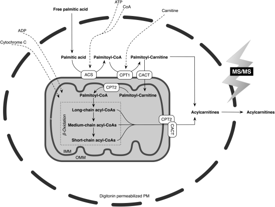
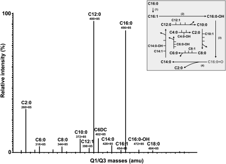
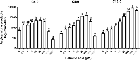
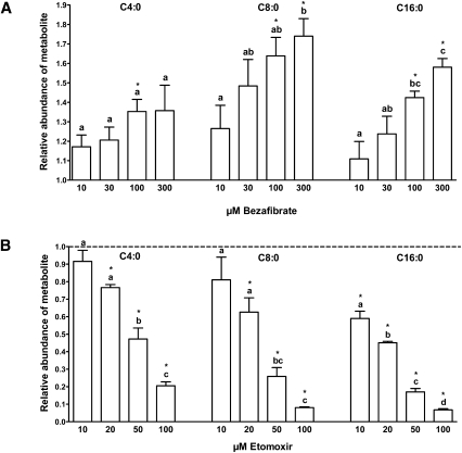
Quantitative analysis of mitochondrial FA β-oxidation (FAO) has drawn increasing interest for defining lipid-induced metabolic dysfunctions, such as in obesity-induced insulin resistance, and evaluating pharmacologic strategies to improve β-oxidation function. The aim was to develop a new assay to quantify β-oxidation function in intact mitochondria and with a low amount of cell material. Cell membranes of primary human fibroblasts were permeabilized with digitonin prior to a load with FFA substrate. Following 120 min of incubation, the various generated acylcarnitines were extracted from both cells and incubation medium by protein precipitation/desalting and subjected to solid-phase extraction. A panel of 30 acylcarnitines per well was quantified by MS/MS and normalized to citrate synthase activity to analyze mitochondrial metabolite flux. Pretreatment with bezafibrate and etomoxir revealed stimulating and inhibiting regulatory effects on β-oxidation function, respectively. In addition to the advantage of a much shorter assay time due to in situ permeabilization compared with whole-cell incubation systems, the method allows the detection of multiple acylcarnitines from an only limited amount of intact cells, particularly relevant to the use of primary cells. This novel approach facilitates highly sensitive, simple, and fast monitoring of pharmacological effects on FAO.


Figures

Fig. 1.
Fig. 1.
Schematic representation of the in situ permeabilization assay for metabolite profiling of palmitate β-oxidation in intact mitochondria. ACS, acyl-CoA synthetase; CACT, carnitine acylcarnitine translocase (officially named as SLC25A20); CPT1, carnitine palmitoyltransferase 1; CPT2, carnitine palmitoyltransferase 2; IMM, inner mitochondrial membrane; OMM, outer mitochondrial membrane; PM, plasma membrane.
Fig. 2.
Fig. 2.
Multiple reaction monitoring (MRM) transitions of acylcarnitines derivatized as their butyl esters following loading with palmitic acid in the in situ permeabilization metabolite assay. Primary human fibroblasts were loaded with palmitic acid (100 µM, 120 min). The signals represent molecular ions of acylcarnitine butyl ester derivatives detected by applying ESI-MS/MS in the MRM mode. Quadrupole Q1/Q3 transitions were monitored by the acylcarnitine-specific daughter ion of 85 amu. The inserted scheme shows the metabolites of mitochondrial palmitic acid β-oxidation, as detected by the in situ permeabilization ESI-MS/MS method. Four enzymatic reactions are involved in each β-oxidation cycle, resulting in consecutive dehydrogenation (1), hydration (2), dehydrogenation (3), and thiolytic cleavage (4) reactions, to generate acetyl-CoA (C2:0) and a new acyl-CoA of two less carbon atoms than the original one. C16:0 = O indicated in gray is not part of the analyzed acylcarnitine panel but is displayed in order to indicate one complete β-oxidation cycle.
Fig. 3.
Fig. 3.
Concentration profile of various acylcarnitine products of palmitate oxidation, analyzed by the in situ permeabilization metabolite assay. Primary human fibroblasts were loaded with eight different concentrations of palmitic acid or incubated with control medium containing no palmitic acid (0 µM) for 120 min. Representative acylcarnitine products of short- (C4:0), medium- (C8:0), and long-chain (C16:0) species are depicted. Three independent experiments were performed in triplicates each. Results are presented as mean ± SEM. For each metabolite, values not sharing a common letter are significantly different (P < 0.05).
Fig. 4.
Fig. 4.
Pharmacological treatment with the PPAR agonist bezafibrate and the CPT1 inhibitor etomoxir followed by palmitate loading. Representative acylcarnitine products of short- (C4:0), medium- (C8:0), and long-chain (C16:0) species are depicted. Three independent experiments were performed in triplicates each. Results are presented as mean ± SEM. Statistically significant differences of pharmacological treatments relative to the untreated control are indicated by an asterisk (P < 0.05). For each metabolite, values not sharing a common letter indicate statistically significant differences in metabolite abundance between concentrations (P < 0.05). A: Stimulation of FA β-oxidation following pretreatment with the PPAR agonist bezafibrate and subsequent palmitate loading (100 µM, 120 min). Bezafibrate was dissolved in DMSO; the final concentration of DMSO per well was 0.05%. Primary human fibroblasts (105 cells/well) were incubated with increasing bezafibrate concentrations (10, 30, 100, 300 µM) for 48 h (38) in DMEM (1 g/l glucose) and 0.25% FA-free BSA. Results are expressed as fold increase in metabolite abundance compared with the untreated control (0.05% DMSO). B: Inhibition of FA β-oxidation following pretreatment with the CPT1 inhibitor etomoxir and subsequent palmitate loading (100 µM, 120 min). Etomoxir was dissolved in H2O. Prior to permeabilization, fibroblasts (105 cells/well) were pretreated with different etomoxir concentrations (10, 20, 50, 100 µM) for 30 min (42) in DMEM (1 g/l glucose) and 0.25% FA-free BSA. Results are expressed as fold decrease in metabolite abundance compared with the untreated control (H2O; indicated by the dotted line).

References

    1. Eaton S., Bartlett K., Pourfarzam M. 1996. Mammalian mitochondrial beta-oxidation. Biochem. J. 320: 345–357 - PMC - PubMed
    1. Matern D., Cuthbert C. D., Tortorelli S., Rinaldo P.2008. Inherited abnormalities in mitochondrial fatty acid oxidation. In Walker's Pediatric Gastrointestinal Disease. R. E. Kleinman, O. J. Goulet, G. M. Vergani, et al., editors. BC Decker, Inc., Hamilton, Ontario. 1287–1307.
    1. Muoio D. M., Newgard C. B. 2008. Mechanisms of disease: molecular and metabolic mechanisms of insulin resistance and beta-cell failure in type 2 diabetes. Nat. Rev. Mol. Cell Biol. 9: 193–205 - PubMed
    1. Koves T. R., Ussher J. R., Noland R. C., Slentz D., Mosedale M., Ilkayeva O., Bain J., Stevens R., Dyck J. R., Newgard C. B., et al. 2008. Mitochondrial overload and incomplete fatty acid oxidation contribute to skeletal muscle insulin resistance. Cell Metab. 7: 45–56 - PubMed
    1. Kølvraa S., Gregersen N., Christensen E., Hobolth N. 1982. In vitro fibroblast studies in a patient with C6-C10-dicarboxylic aciduria: evidence for a defect in general acyl-CoA dehydrogenase. Clin. Chim. Acta. 126: 53–67 - PubMed

Publication types

LinkOut - more resources