

An unusual case of Takayasu's arteritis: Evaluation by CT angiography
- PMID: 22346024
- PMCID: PMC3271474
- DOI: 10.4103/0972-2327.91960
An unusual case of Takayasu's arteritis: Evaluation by CT angiography
Abstract
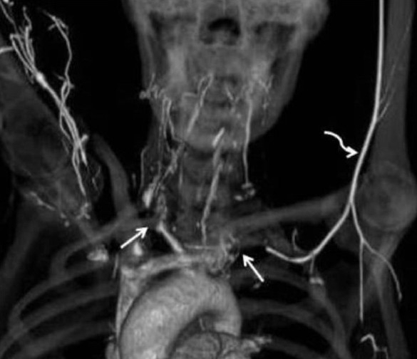
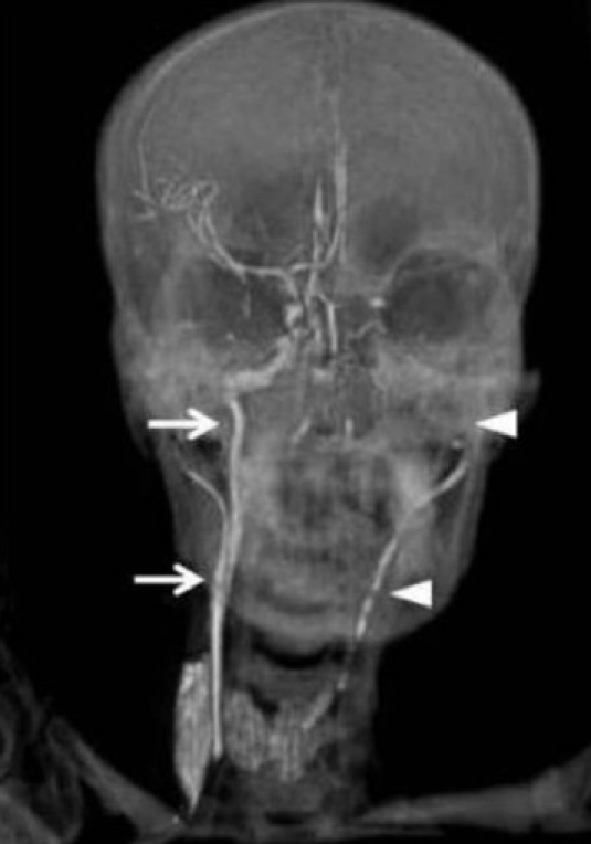
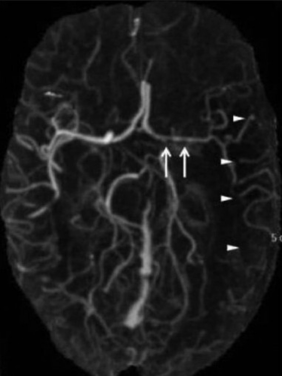
Takayasu's arteritis is a chronic, idiopathic, medium and large vessel vasculitis involving aorta and its main branches. Frequent neurological manifestations include postural syncope, seizures, and blindness. Stroke, as presenting feature of Takayasu's arteritis, is unusual. CT angiography reveals characteristic involvement of aortic arch and its branches. Involvement of intracranial vasculature is rather unusual. We are describing an unusual patient of Takayasu's arteritis who presented with recurrent disabling syncopal attacks and had extensive involvement of intracranial vasculature. CT angiography revealed severe involvement of aortic arch. There was near complete occlusion at origins of both subclavian arteries, distal flow was maintained by collateral vessels along the chest wall. There was near total occlusion (at origin) of right common carotid with normal flow in distal part. The left common carotid was more severely involved showing greater than 80% narrowing in proximal half of the vessel. CT angiography also revealed involvement of left internal carotid artery, narrowing of left middle cerebral artery and involvement of cortical vessels. Patient was treated with oral corticosteroids. She improved remarkably after two and half months of follow up.
Keywords: Takayasu's arteritis; stroke; vasculitis.
Conflict of interest statement
Figures
Similar articles
-
Ultrasonographic study and long-term follow-up of Takayasu's arteritis.Stroke. 1996 Dec;27(12):2178-82. doi: 10.1161/01.str.27.12.2178. Stroke. 1996. PMID: 8969776
-
Bilateral Takayasu's retinopathy as the initial presentation of Takayasu's arteritis.BMJ Case Rep. 2024 Apr 29;17(4):e258688. doi: 10.1136/bcr-2023-258688. BMJ Case Rep. 2024. PMID: 38684359
-
Subclavian artery aneurysm: an unusual manifestation of Takayasu's arteritis.Cardiovasc Surg. 1999 Apr;7(3):310-4. doi: 10.1016/s0967-2109(98)00171-9. Cardiovasc Surg. 1999. PMID: 10386748
-
Small vessel involvement in Takayasu's arteritis.Autoimmun Rev. 2013 Jan;12(3):355-62. doi: 10.1016/j.autrev.2012.05.010. Epub 2012 Jun 9. Autoimmun Rev. 2013. PMID: 22691438 Review.
-
The Epidemiology and Clinical Manifestations of Takayasu Arteritis: A Descriptive Study of Case Reports.Cureus. 2021 Sep 15;13(9):e17998. doi: 10.7759/cureus.17998. eCollection 2021 Sep. Cureus. 2021. PMID: 34667674 Free PMC article. Review.
Cited by
-
Takayasu Arteritis Presenting as Bilateral Ocular Ischemic Syndrome.Vasc Specialist Int. 2020 Sep 30;36(3):163-169. doi: 10.5758/vsi.200031. Vasc Specialist Int. 2020. PMID: 32868487 Free PMC article.
-
Bilateral ocular ischemia-induced blindness as a presenting manifestation of Takayasu arteritis: a case report.J Med Case Rep. 2017 Jun 10;11(1):153. doi: 10.1186/s13256-017-1330-3. J Med Case Rep. 2017. PMID: 28599682 Free PMC article.
-
Vascular Imaging Techniques to Diagnose and Monitor Patients with Takayasu Arteritis: A Review of the Literature.Diagnostics (Basel). 2021 Oct 27;11(11):1993. doi: 10.3390/diagnostics11111993. Diagnostics (Basel). 2021. PMID: 34829340 Free PMC article. Review.
-
Multiple occlusions in extracranial arteries in patients with aortic arch syndrome: is minimally invasive treatment still possible? Technical aspects of the treatment based on our own experience and a review of the literature.Wideochir Inne Tech Maloinwazyjne. 2021 Mar;16(1):183-190. doi: 10.5114/wiitm.2020.94517. Epub 2020 Apr 20. Wideochir Inne Tech Maloinwazyjne. 2021. PMID: 33786133 Free PMC article.
References
-
- Kerr GS, Hallahan CW, Giordano J, Leavitt RY, Fauci AS, Rottem M, et al. Takayasu's Arteritis. Ann Intern Med. 1994;120:919–29. - PubMed
-
- Khandelwal N, Kalra N, Garg MK, Kanga M, Lal A, Jain S, et al. Multidetector CT angiography in Takayasu arteritis. Eur J Radiol. 2011;77:369–74. - PubMed
-
- Sikaroodi H, Motamedi M, Kahnooji H, Gholamrezanezhad A, Yousefi N. Stroke as the first manifestation of Takayasu arteritis. Acta Neurol Belg. 2007;107:18–21. - PubMed
-
- Klos K, Flemming KD, Petty GW, Luthra HS. Takayasu's arteritis with arteriographic evidence of intracranial vessel involvement. Neurology. 2003;60:1550–1. - PubMed
