

A case of leukoencephalopathy, cerebral calcifications and cysts
- PMID: 22346026
- PMCID: PMC3271476
- DOI: 10.4103/0972-2327.91964
A case of leukoencephalopathy, cerebral calcifications and cysts
Abstract
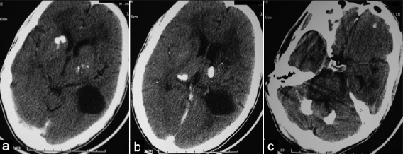
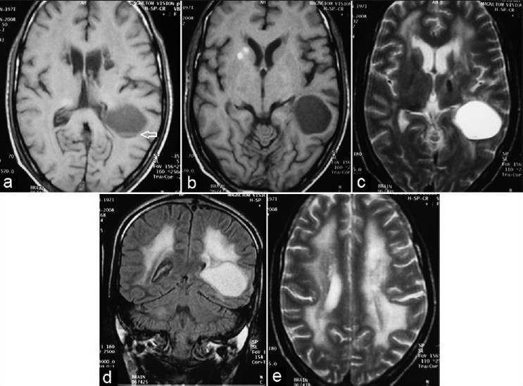
Triad of leukoencephalopathy, cerebral calcifications and cysts (LCC) is a recently reported rare disease named 'Labrune syndrome' after the first case was reported in 1996 by Labrune et al. Herein, we report a case of a 36-year-old man with mild right-sided weakness and seizures for 5 years. CT of brain revealed extensive calcification involving bilateral basal ganglia, right thalamus and bilateral deep cerebellar nuclei. A supratentorial cystic lesion with blood fluid level was seen in left occipitotemporal region. MRI examination revealed diffuse symmetric white matter hyperintensity suggesting leukoencephalopathy. On follow-up, patient reported improvement in the weakness and no further seizure episodes. However, follow-up of MRI revealed persistence of lesions. Differential diagnosis considered were parasitic infections (hydatid, cysticercosis), Coat's plus disease and causes of diffuse cerebral calcification like Fahr's disease and post-radiotherapy/chemotherapy. Serology for parasitic infections was negative. No history of radiotherapy or chemotherapy in the past could be elicited in the history. Another close differential is Coat's plus disease which can mimic LCC pathologically.
Keywords: Calcification; cerebral calcifications and cysts; cerebral cysts; labrune syndrome; leukoencephalopathy.
Conflict of interest statement
Figures
References
-
- Labrune P, Lacroix C, Goutieres F, de Laveaucoupet J, Chevalier P, Zerah M, et al. Extensive brain calcifications, leukodystrophy, and formation of parenchymal cysts: A new progressive disorder due to diffuse cerebral microangiopathy. Neurology. 1996;46:1297–301. - PubMed
-
- Nagae-Poetscher LM, Bibat G, Philippart M, Rosemberg S, Fatemi A, Lacerda MT, et al. Leukoencephalopathy, cerebral calcifications, and cysts: New observations. Neurology. 2004;62:1206–9. - PubMed
-
- Turkulov V, Madle-Samardzija N, Canak G, Vukadinov J, Aleksic-Dordevic M. Clinical and diagnostic approaches to neurocysticercosis. Med Pregl. 2001;54:353–6. - PubMed
-
- Corboy JR, Gault J, Kleinschmidt-DeMasters BK. An adult case of leukoencephalopathy with intracranial calcifications and cysts. Neurology. 2006;67:1890–2. - PubMed
Publication types
LinkOut - more resources
Full Text Sources
Miscellaneous
