Interceptive orthopedics for the correction of maxillary transverse and sagittal deficiency in the early mixed dentition period
- PMID: 22346162
- PMCID: PMC3276862
- DOI: 10.4103/0976-237X.91798
Interceptive orthopedics for the correction of maxillary transverse and sagittal deficiency in the early mixed dentition period
Abstract
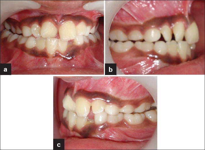
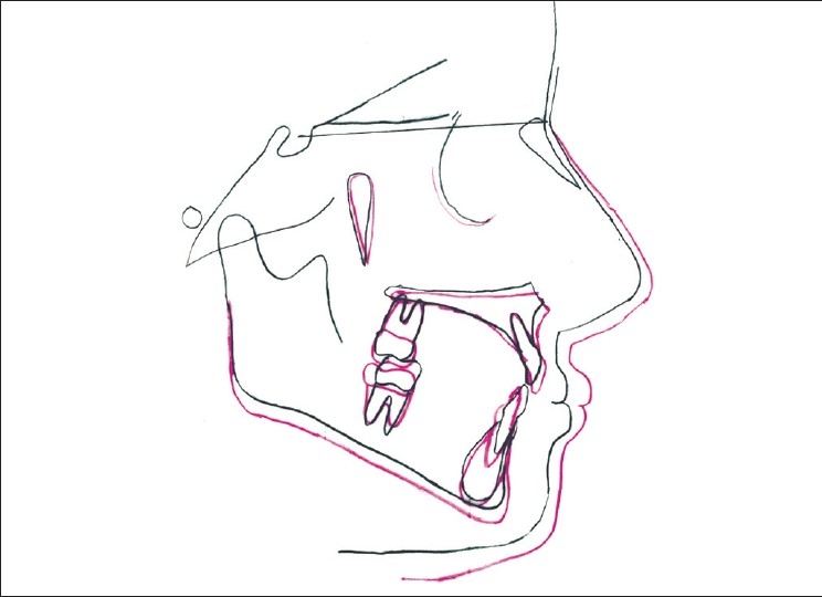
Dentofacial Orthopedics directed to a hypoplastic maxilla in the prepubertal period redirects growth of the maxilla in the vertical, transverse and sagittal planes of space. The orthopedic correction of maxillary hypoplasia in the early mixed dentition period thus intercepts the establishment of permanent structural asymmetry in the mandible and helps in the achievement of optimal dentofacial esthetics. This paper presents the growth redirection in a hypoplastic maxilla of an 8-year-old girl with simultaneous rapid maxillary expansion and protraction headgear therapy for a period of 11 months which corrected the posterior unilateral cross-bite, the positional asymmetry of the mandible and established an orthognathic profile in the individual.
Keywords: Face mask appliance; mandibular structural asymmetry; posterior unilateral cross-bite; rapid maxillary expansion.
Conflict of interest statement
Figures


Similar articles
-
Soft tissue and dentoskeletal profile changes associated with maxillary expansion and protraction headgear treatment.Am J Orthod Dentofacial Orthop. 1996 Jan;109(1):38-49. doi: 10.1016/s0889-5406(96)70161-0. Am J Orthod Dentofacial Orthop. 1996. PMID: 8540481
-
Intercanine widening and sagittal effect of maxillary transverse expansion in patients with cleft lip and palate during the deciduous and mixed dentitions.Cleft Palate Craniofac J. 1993 Mar;30(2):195-207. doi: 10.1597/1545-1569_1993_030_0195_iwaseo_2.3.co_2. Cleft Palate Craniofac J. 1993. PMID: 8452842
-
Soft-tissue profile changes during widening and protraction of the maxilla in patients with cleft lip and palate compared with normal growth and development.Cleft Palate Craniofac J. 1993 Sep;30(5):454-68. doi: 10.1597/1545-1569_1993_030_0454_stpcdw_2.3.co_2. Cleft Palate Craniofac J. 1993. PMID: 8218309
-
Realities of craniofacial growth modification.Atlas Oral Maxillofac Surg Clin North Am. 2001 Mar;9(1):23-51. Atlas Oral Maxillofac Surg Clin North Am. 2001. PMID: 11905336 Review.
-
Clinical effects of maxillary protraction in different stages of dentition in skeletal class III children: A systematic review and meta-analysis.Orthod Craniofac Res. 2022 Nov;25(4):549-561. doi: 10.1111/ocr.12569. Epub 2022 Mar 23. Orthod Craniofac Res. 2022. PMID: 35303382
References
-
- Arman A, Ufuk Toygar T, Abuhijleh E. Profile changes associated with different orthopedic treatment approaches in class III malocclusions. Angle Orthod. 2004;74:734–40. - PubMed
-
- Irie M, Nakamura S. Orthopedic approach to severe class III malocclusion. Am J Orthod. 1975;67:337–92. - PubMed
-
- Cozzani G. Extraoral traction and class ill treatment. Am J Orthod. 1981;80:638–50. - PubMed
-
- Guyer EC, Ellis EE, 3rd, McNamara JA, Jr, Behrents RG. Components of class III malocclusion in juveniles and adolescents. Angle Orthod. 1986;56:7–30. - PubMed
-
- Kerr WJ, Ten Hane TR. Comparison of three appliance systems in the treatment of class III malocclusion. Eur J Orthod. 1988;10:203–14. - PubMed