Solitary median maxillary central incisor in association with hemifacial microsomia: A rare case report and review of literature
- PMID: 22346174
- PMCID: PMC3276874
- DOI: 10.4103/0976-237X.91810
Solitary median maxillary central incisor in association with hemifacial microsomia: A rare case report and review of literature
Abstract
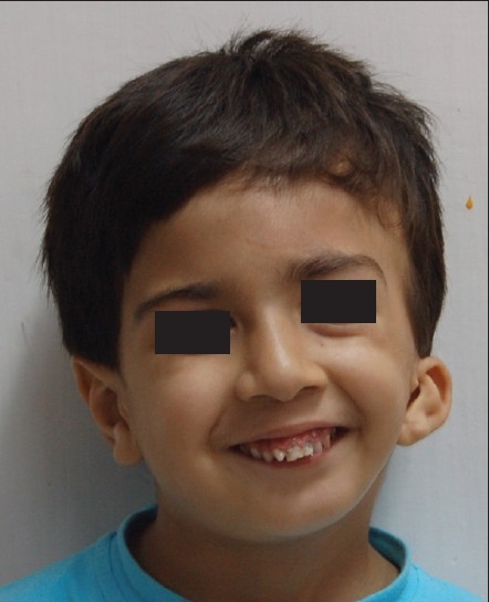
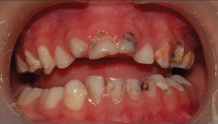
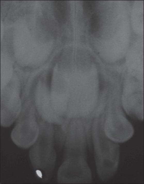
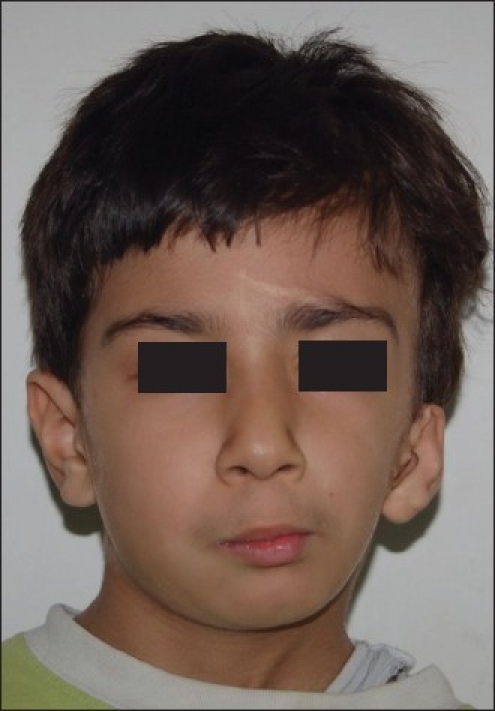
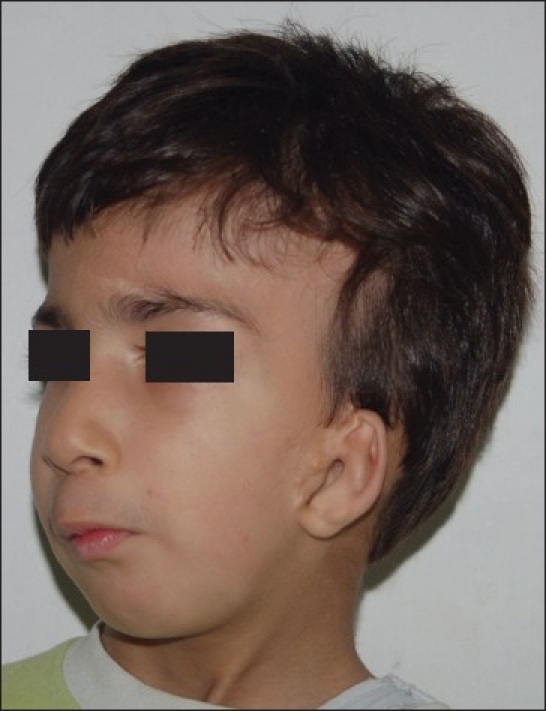
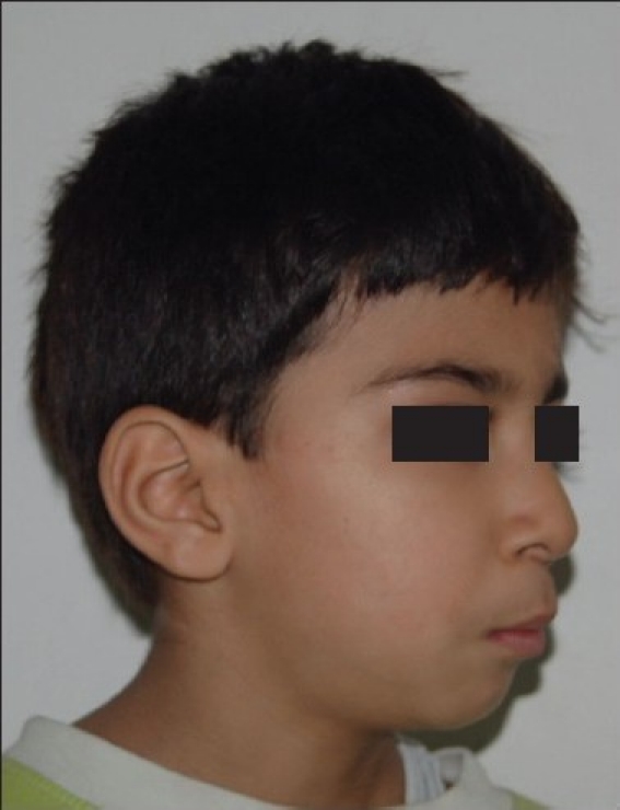
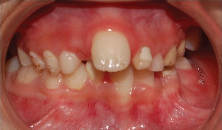
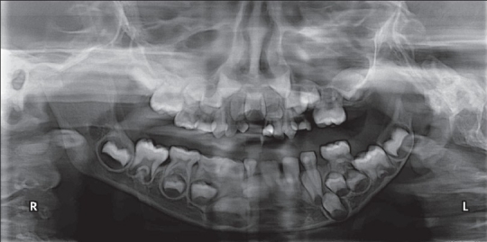
Solitary median maxillary central incisor (SMMCI) is a rare dental anomaly. It is estimated to occur in 1:50,000 live births. The SMMCI tooth differs from the normal central incisor in that the crown form is symmetric and it develops and erupts precisely in the midline of the maxillary dental arch in both primary and permanent dentitions. Presence of SMMCI with hemifacial microsomia (HFM) is a very rare clinical condition. We report a case of HFM in a male of Indian origin who presented with SMMCI in both primary and permanent dentitions. The association of HFM with SMMCI may be due to defective development of neural crest cells and/or lack of space in maxilla.
Keywords: Hemifacial microsomia; anodontia; solitary median maxillary central incisor.
Conflict of interest statement
Figures
References
-
- Garavelli L, Zanacca C, Caselli G, Banchini G, Dubourg C, David V, et al. Solitary median maxillary central incisor syndrome: clinical case with a novel mutation of sonic hedgehog. Am J Med Genet A. 2004;127:93–5. - PubMed
-
- Scott DC. Absence of upper central incisor. Br Dent J. 1958;104:247–8.
-
- Hall RK, Bankier A, Aldred MJ, Kan K, Lucas JO, Perks AG. Solitary median maxillary central incisor, short stature, choanal atresia/midnasal stenosis (SMMCI) syndrome. Oral Surg Oral Med Oral Pathol Oral Radiol Endod. 1997;84:651–62. - PubMed
-
- Rappaport EB, Ulstrom R, Gorlin RJ. Monosuperocentroinci-sivodontic dwarfism. Birth Defects Orig Artic Ser. 1976;12:243–5. - PubMed
-
- Maréchaux SC. The single maxillary central primary incisor: report of case. ASDC J Dent Child. 1986;53:124–12. - PubMed
Publication types
LinkOut - more resources
Full Text Sources
Molecular Biology Databases