. 2012:7:547-57.
doi: 10.2147/IJN.S26141. Epub 2012 Feb 2.

Preventing postoperative abdominal adhesions in a rat model with PEG-PCL-PEG hydrogel

Affiliations

Preventing postoperative abdominal adhesions in a rat model with PEG-PCL-PEG hydrogel

Bing Yang et al. Int J Nanomedicine. 2012.

Abstract

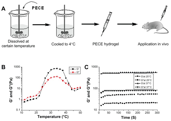
Background: Poly (ethylene glycol)-poly (ɛ-caprolactone)-poly (ethylene glycol) (PEG-PCL-PEG, PECE) hydrogel has been demonstrated to be biocompatible and thermosensitive. In this study, its potential efficacy and mechanisms of preventing postsurgical abdominal adhesions were investigated.

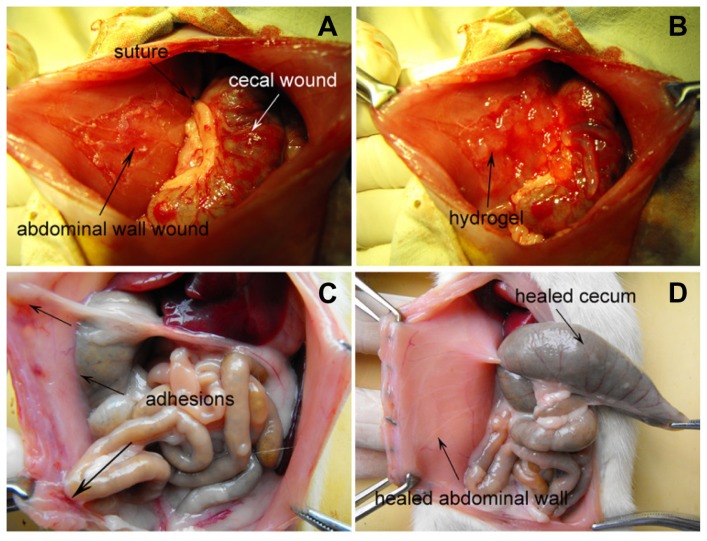
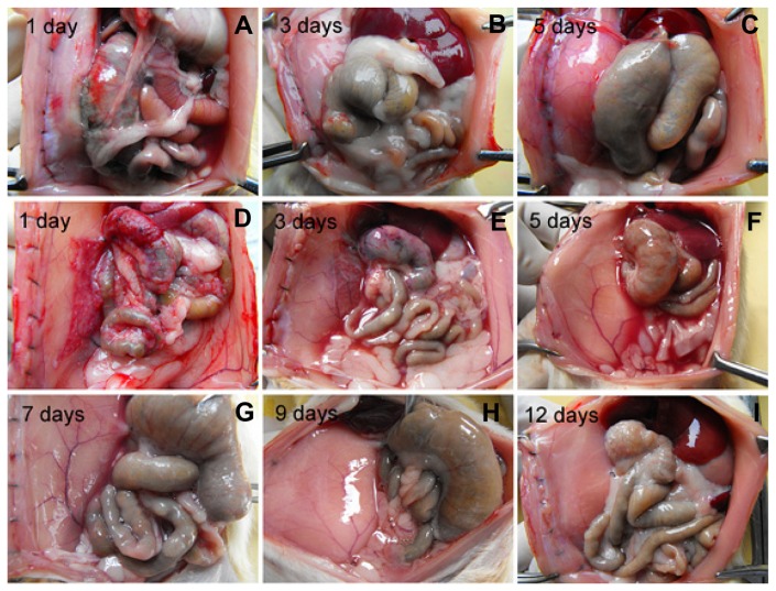
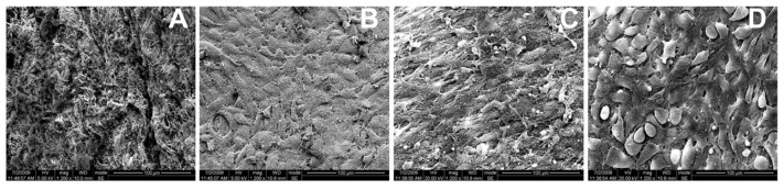
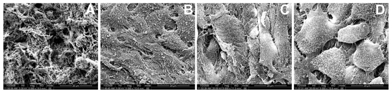
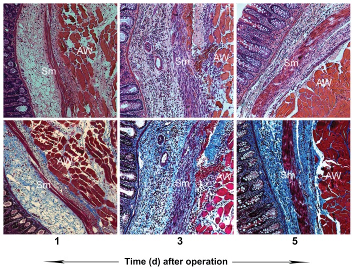
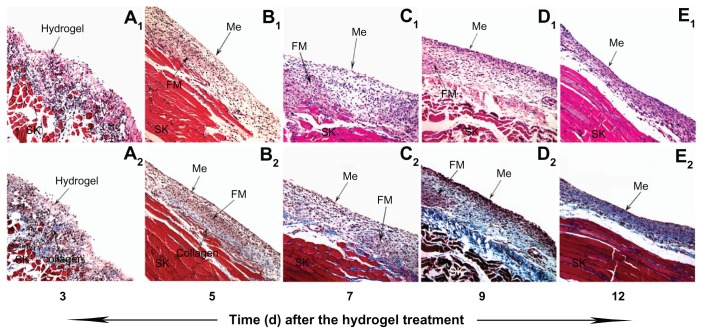
Results: PECE hydrogel was transformed into gel state from sol state in less than 20 seconds at 37°C. None of the animals treated with the hydrogel (n = 15) developed adhesions. In contrast, all untreated animals (n = 15) had adhesions that could only be separated by sharp dissection (P < 0.001). The hydrogel adhered to the peritoneal wounds, gradually disappeared from the wounds within 7 days, and transformed into viscous fluid, being completely absorbed within 12 days. The parietal and visceral peritoneum were remesothelialized in about 5 and 9 days, respectively. The hydrogel prevented the formation of fibrinous adhesion and the invasion of fibroblasts. Also, along with the hydrogel degradation, a temporary inflammatory cell barrier was formed which could effectively delay the invasion of fibroblasts during the critical period of mesothelial regeneration.

Conclusion: The results suggested that PECE hydrogel could effectively prevent postsurgical intra-abdominal adhesions, which possibly result from the prevention of the fibrinous adhesion formation and the fibroblast invasion, the promotion of the remesothelialization, and the hydroflotation effect.

Keywords: anti-adhesion; barrier; biocompatible; thermosensitive.


Figures

Figure 1
Figure 1
(A) Schematic illustration of PECE hydrogel for in vivo application. (B) Rheology analysis of PECE hydrogel as a function of temperature. (C) Time dependence of storage modulus G′ and loss modulus G″ at different temperatures. Abbreviation: PECE, poly (ethylene glycol)-poly (ɛ-caprolactone)-poly (ethylene glycol) (PEG-PCL-PEG).
Figure 2
Figure 2
(A) The rat model of abdominal sidewall defect-cecum abrasion. (B) PECE hydrogel applied on the injured abdominal wall and cecum. (C) Score-4 adhesion was observed between the injured abdominal wall and cecum in an NS-treated rat; intestine segments and omentum adhesion to the sutured midline incisions were also observed. (D) No adhesion was observed in the PECE gel-treated group. Abbreviations: NS, normal saline; PECE, poly (ethylene glycol)-poly (ɛ-caprolactone)-poly (ethylene glycol) (PEG-PCL-PEG).
Figure 3
Figure 3
Autopsy observation in NS-treated group and hydrogel-treated group at different times. (A, B, and C) Adhesion formation on the indicated days in the NS group. (D, E, F, G, H, and I) The degradation of the adhered hydrogel and healing of the injured surfaces on the indicated days in the treatment group. Abbreviation: NS, normal saline.
Figure 4
Figure 4
Scanning electron microscopy image of the surface of the stripped peritoneum. (A) The injured surface was covered by a layer of hydrogel on day 3. (B) The surface was covered with a layer of elongated, flattened, squamous-shaped cells on day 5. The squamous-shaped cells junction became tight on day 7 (C) and loose again on day 9 (D).
Figure 5
Figure 5
Scanning electron microscopy image of the surface of the stripped peritoneum (higher magnification of Figure 4). (A) The hydrogel covered the meshwork of fibrin, and several small spherical inflammatory cells were observed at day 3. (B) Large squamous-shaped cells over the surface and these cells had short microvilli. These microvilli increased and elongated at days 7 (C) and 9 (D).
Figure 6
Figure 6
Postsurgical adhesion formation at different times in rats treated with NS. Note that, along with days after the operation, infiltration with inflammatory cells (upper) and collagen deposition (lower) in both abdomen walls and cecal walls increased. Abbreviations: AW, abdominal wall muscle; NS, normal saline; Sm, smooth muscle.
Figure 7
Figure 7
The fibrosis and the remesothelialization of the peritoneal wounds treated with PECE hydrogel. Three days after the treatment, a coating of the hydrogel on the injured surface of the abdominal wall, with infiltration of inflammatory cells (A1, 200×) and scattered collagen beneath (A2, 200×). Five days after the treatment, the layer of inflammatory cells composed mainly of foamy macrophages appeared, above which was a layer of spindle-shaped mesothelial cells (B1, 200×), and under which was a collagen layer (B2, 200×). Along with days, collagen- and fibroblast-rich tissues gradually increased and replaced the structure of the inflammatory cell layer (C1, C2, D1, D2, E1, E2, 200×). Abbreviations: Me, mesothelial cell layer; SK, skeletal muscle; FM, foamy macrophages; PECE, poly (ethylene glycol)-poly (ɛ-caprolactone)-poly (ethylene glycol) (PEG-PCL-PEG).

Similar articles

Cited by

References

    1. Weibel MA, Majno G. Peritoneal adhesions and their relation to abdominal surgery. A postmortem study. Am J Surg. 1973;126:345–353. - PubMed
    1. Menzies D, Ellis H. Intestinal obstruction from adhesions – how big is the problem. Ann R Coll Surg Engl. 1990;72:60–63. - PMC - PubMed
    1. Group OLS. Postoperative adhesion development after operative laparoscopy: evaluation at early second-look procedures. Operative Laparoscopy Study Group. Fertil Steril. 1991;55:700–704. - PubMed
    1. Best CL, Rittenhouse D, Vasquez C, Norng T, Subias E, Sueldo CE. Evaluation of interceed(TC7) for reduction of postoperative adhesions in rabbits. Fertil Steril. 1992;58:817–820. - PubMed
    1. DeCherney AH, diZerega GS. Clinical problem of intraperitoneal post-surgical adhesion formation following general surgery and the use of adhesion prevention barriers. Surg Clin North Am. 1997;77:671–688. - PubMed

Publication types

MeSH terms

Substances