. 2012 Feb;8(2):e1002369.
doi: 10.1371/journal.pcbi.1002369. Epub 2012 Feb 9.

Control of whole heart geometry by intramyocardial mechano-feedback: a model study

Affiliations

Control of whole heart geometry by intramyocardial mechano-feedback: a model study

Theo Arts et al. PLoS Comput Biol. 2012 Feb.

Abstract

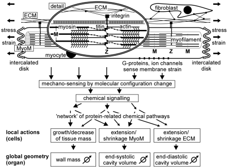
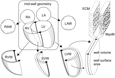
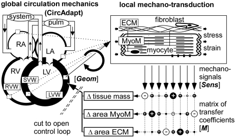
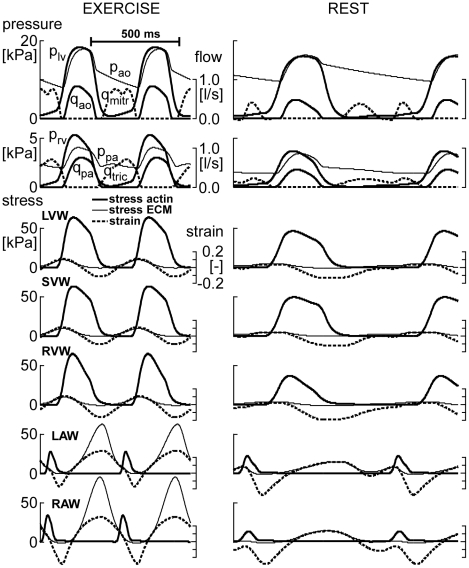
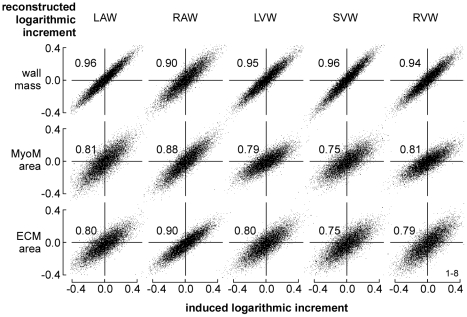
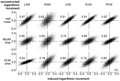
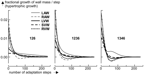
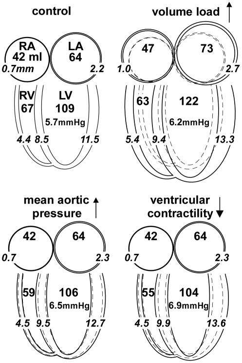
Geometry of the heart adapts to mechanical load, imposed by pressures and volumes of the cavities. We regarded preservation of cardiac geometry as a homeostatic control system. The control loop was simulated by a chain of models, starting with geometry of the cardiac walls, sequentially simulating circulation hemodynamics, myofiber stress and strain in the walls, transfer of mechano-sensed signals to structural changes of the myocardium, and finalized by calculation of resulting changes in cardiac wall geometry. Instead of modeling detailed mechano-transductive pathways and their interconnections, we used principles of control theory to find optimal transfer functions, representing the overall biological responses to mechanical signals. As biological responses we regarded tissue mass, extent of contractile myocyte structure and extent of the extra-cellular matrix. Mechano-structural stimulus-response characteristics were considered to be the same for atrial and ventricular tissue. Simulation of adaptation to self-generated hemodynamic load rendered physiologic geometry of all cardiac cavities automatically. Adaptation of geometry to chronic hypertension and volume load appeared also physiologic. Different combinations of mechano-sensors satisfied the condition that control of geometry is stable. Thus, we expect that for various species, evolution may have selected different solutions for mechano-adaptation.


Conflict of interest statement

The authors have declared that no competing interests exist.

Figures

Figure 1
Figure 1. Schematic of adaptation of wall geometry to mechanical load.
Global stress and strain of the tissue determine forces and deformation of the myocyte matrix (MyoM) and extracellular matrix (ECM). Within the myocyte, axial forces are born by the sarcomeres in the myofilaments, consisting of actin and myosin, mutually coupled by cross-bridges. The Z-disk connects actin and titin of neighboring sarcomeres. The distance between Z-discs represents sarcomere length, ranging from 1.7 to 2.3 µm. Titin parallels actin, and is connected to myosin. The Z-disks are connected transversely to the ECM by integrins, traversing the cell membrane of the myocyte. Mechano-sensing has been reported around the Z-disk, within the cell membrane of the myocyte and in the intercalated disks, forming axial connections between myocytes. Fibroblasts are also mechano-sensitive. Resulting chemical signals follow a network of intertwined pathways of chemical activity, resulting in local actions of adaptation. Summed effects of local actions determine global changes in geometry of the myocardial wall.
Figure 2
Figure 2. Composition of the heart by 5 walls with MyoM and ECM.
Curved mid-wall surface form the core of macroscopic geometry of atria and ventricles. Left and right atrium (LA, RA) are enclosed by single curved walls (LAW, RAW). The ventricular unit is composed of left, septal and right ventricular wall (LVW, SVW and RVW), enclosing left and right ventricle (LV, RV). Geometry of each wall is deducted from unfolding the wall to a flat surface. Each wall has a volume and a mid-wall area. A wall contains a myocyte matrix (MyoM) intertwined with the extracellular matrix (ECM). Both matrices have their own reference mid-wall area, linking their ultrastructure to the macroscopic geometry of the wall.
Figure 3
Figure 3. Modeling control of myocardial wall geometry.
In the CircAdapt model of the whole circulation (left), geometry of the walls determines hemodynamic performance, which in turn determines mechanical load of the wall material (upper right). Local mechano-sensing invokes changes in tissue structure (lower mid) and geometry, by which the control loop for adaptation of wall geometry is closed. A matrix of transfer coefficients (lower right) is used to estimate changes in tissue structure, causing geometry to adapt. Symbols [Sens, M, Geom] are used in Eq. (2–9). Abbreviations are explained in the general list.
Figure 4
Figure 4. Simulation of circulation dynamics and cardiomechanics.
With the CircAdapt model, beat-to-beat circulation dynamics are simulated at moderate exercise (left) and at rest (right). Top: Pressures in left/right ventricle (plv/prv) and aorta/pulmonary artery (pao/ppa), and flows through aortic/pulmonary (qao/qpa) and mitral/tricuspid valves (qmitr/qtric). Lower panels: Stresses in actin and ECM of the 5 cardiac walls and related fiber strains. For exercise and rest, amplitude and time calibrations are the same. Abbreviations LAW, RAW, LVW, SVW, RVW are defined in Fig. 2.
Figure 5
Figure 5. Reconstruction of cardiac wall geometry by mechano-sensing.
A large number (10,000) of random increments of structural parameters was induced in all cardiac walls. In each wall, information about mechanical load of the tissue was obtained from 8 locally sensed variables (Table 2). With this information, best estimates were made of the originally induced increments. For the 3 structural parameters (vertical) and 5 walls (horizontal) reconstructed increment is plotted as a function of original increment. Numbers in the graphs indicate correlation coefficients.
Figure 6
Figure 6. Reconstruction of wall geometry from 3 sensed variables (126).
Mechano-sensed variables, used for feedback, are Secm,max, Sact,max, and Ls,int according to Table 2. For explanation, see also Fig. 5.
Figure 7
Figure 7. Simulation of hypertrophic growth of the five myocardial walls.
The fractional change of wall mass per adaptation step is plotted as a function of the number of steps after 20% increase of cardiac output. The numbers 126, 1346 and 1236 refer to the applied combination of sensed variables, used for mechano-feedback (Table 3). After fast mass growth during the first 25 cycles, for combination 126 a slow component remains active for a long time. Convergence with 1236 is best. Convergence with 1346 is also fast, but there is more overshoot.
Figure 8
Figure 8. Adaptation to a 20% change of pump load and contractility.
Mean volumes of right and left atria (RA, LA) and right and left ventricles (RV, LV), respectively, are indicated by the larger numbers in ml. Mean thicknesses of atrial and ventricular walls are indicated by italic numbers in mm. Drawn changes in geometry are exaggerated for better visibility. End-diastolic pressures are indicated in mmHg. With 20% increase of stroke volume, all volumes and thicknesses increase (eccentric hypertrophy), except for RV volume. With 20% increase of mean aortic pressure, walls thicken, while geometry does not change much. With a 20% decrease of LV and septal contractility, these walls thicken, while further geometry is preserved.

References

    1. Ruwhof C, van der Laarse A. Mechanical stress-induced cardiac hypertrophy: mechanisms and signal transduction pathways. Cardiovasc Res. 2000;47:23–37. - PubMed
    1. Lammerding J, Kamm RD, Lee RT. Mechanotransduction in cardiac myocytes. Ann N Y Acad Sci. 2004;1015:53–70. - PubMed
    1. Heineke J, Molkentin JD. Regulation of cardiac hypertrophy by intracellular signalling pathways. Nat Rev Mol Cell Biol. 2006;7:589–600. - PubMed
    1. Linke WA. Sense and stretchability: the role of titin and titin-associated proteins in myocardial stress-sensing and mechanical dysfunction. Cardiovasc Res. 2008;77:637–648. - PubMed
    1. Brancaccio M, Hirsch E, Notte A, Selvetella G, Lembo G, et al. Integrin signalling: the tug-of-war in heart hypertrophy. Cardiovasc Res. 2006;70:422–433. - PubMed

Publication types