. 2012 Feb;8(2):e1002383.
doi: 10.1371/journal.pcbi.1002383. Epub 2012 Feb 9.

Subcellular location of PKA controls striatal plasticity: stochastic simulations in spiny dendrites

Affiliations

Subcellular location of PKA controls striatal plasticity: stochastic simulations in spiny dendrites

Rodrigo F Oliveira et al. PLoS Comput Biol. 2012 Feb.

Abstract

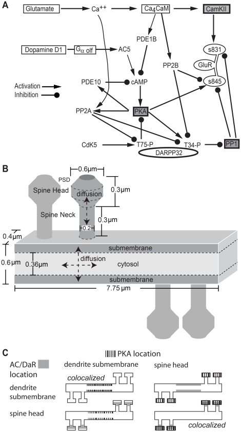
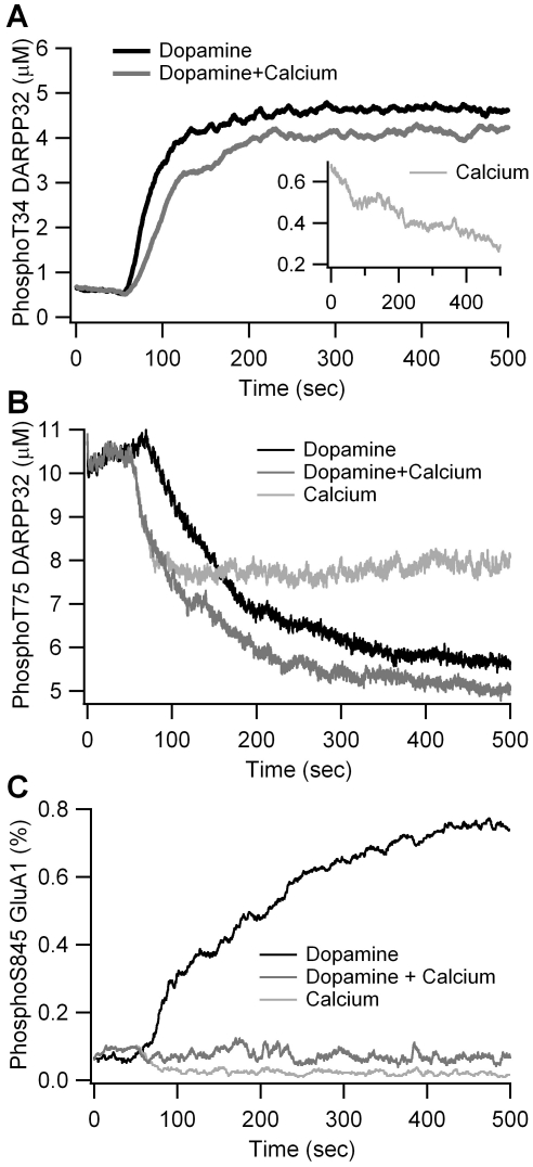
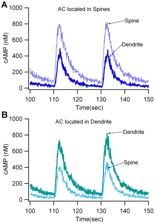
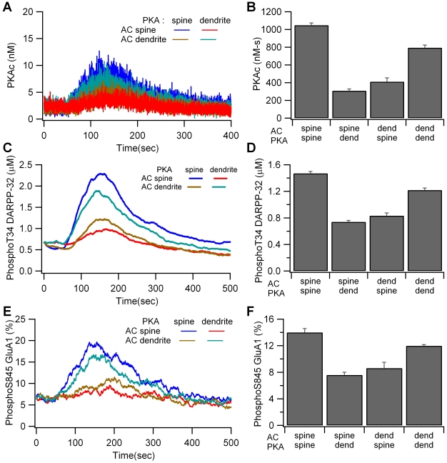
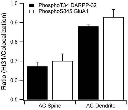
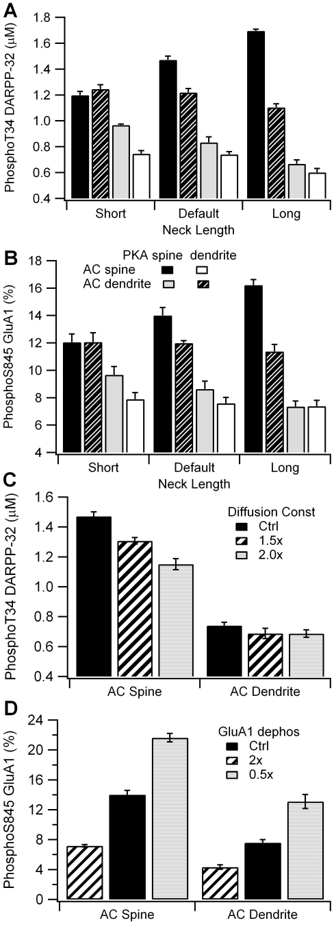
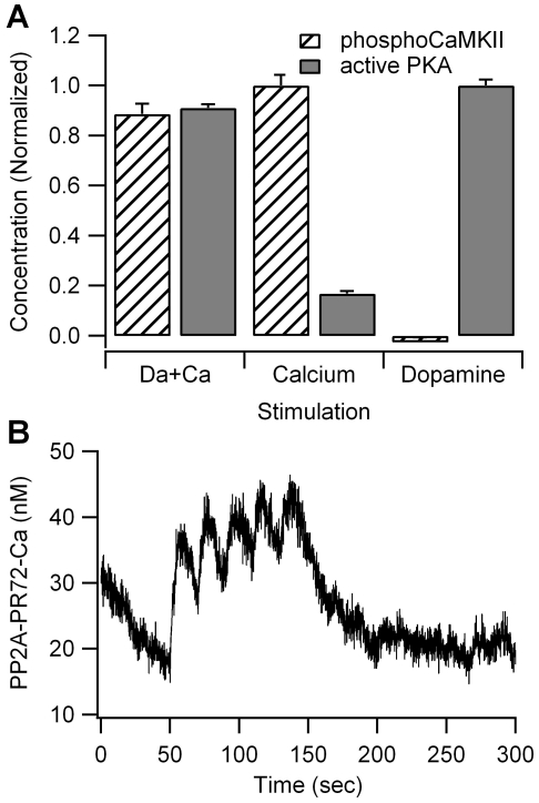
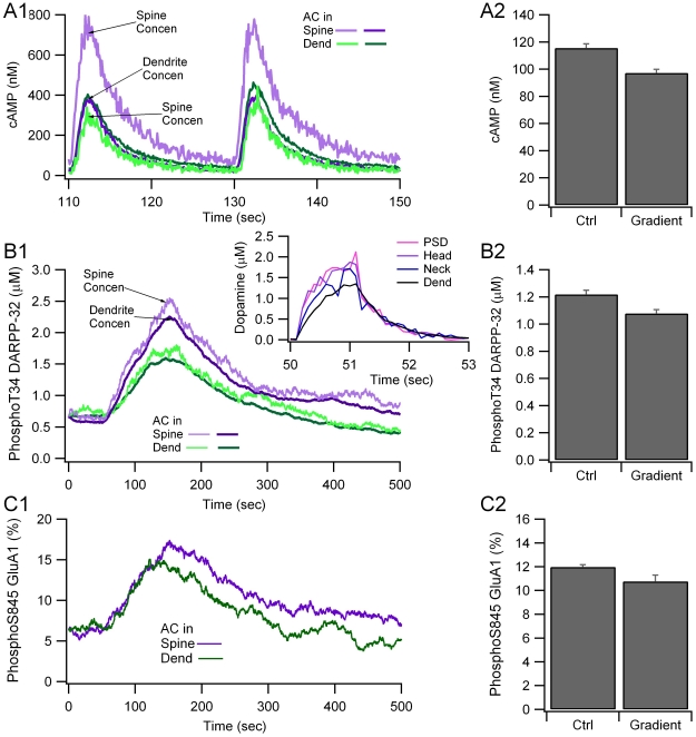
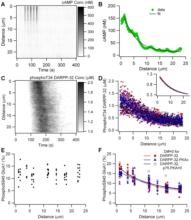
Dopamine release in the striatum has been implicated in various forms of reward dependent learning. Dopamine leads to production of cAMP and activation of protein kinase A (PKA), which are involved in striatal synaptic plasticity and learning. PKA and its protein targets are not diffusely located throughout the neuron, but are confined to various subcellular compartments by anchoring molecules such as A-Kinase Anchoring Proteins (AKAPs). Experiments have shown that blocking the interaction of PKA with AKAPs disrupts its subcellular location and prevents LTP in the hippocampus and striatum; however, these experiments have not revealed whether the critical function of anchoring is to locate PKA near the cAMP that activates it or near its targets, such as AMPA receptors located in the post-synaptic density. We have developed a large scale stochastic reaction-diffusion model of signaling pathways in a medium spiny projection neuron dendrite with spines, based on published biochemical measurements, to investigate this question and to evaluate whether dopamine signaling exhibits spatial specificity post-synaptically. The model was stimulated with dopamine pulses mimicking those recorded in response to reward. Simulations show that PKA colocalization with adenylate cyclase, either in the spine head or in the dendrite, leads to greater phosphorylation of DARPP-32 Thr34 and AMPA receptor GluA1 Ser845 than when PKA is anchored away from adenylate cyclase. Simulations further demonstrate that though cAMP exhibits a strong spatial gradient, diffusible DARPP-32 facilitates the spread of PKA activity, suggesting that additional inactivation mechanisms are required to produce spatial specificity of PKA activity.


Conflict of interest statement

The authors have declared that no competing interests exist.

Figures

Figure 1
Figure 1. Model of striatal medium spiny projection neuron dendrite with spines.
A. Diagram of biochemical signaling pathways. Each arrow is modeled with one or more bimolecular or enzyme reactions. See text and Table 1 for details. B. Morphology of dendrite with four spines, and location of calcium influx in the model. Subvolumes of height 0.12 µm adjacent to the top and bottom surface of the dendrite are considered submembrane subvolumes. Other dendritic subvolumes are part of the cytosol. Diffusion is two-dimensional in the dendrite and one-dimensional in the spine. C. Experimental Design: The role of anchoring is evaluated using four spatial variations in the location of adenylate cyclase and PKA. The adenylate cyclase-D1R complex (AC) is located either in the spine head or a focal dendritic submembrane area. Similarly, the PKA holoenzyme is located either in the spine head or the focal dendritic submembrane area. AMPA receptors containing GluA1 subunits are in the PSD compartment of the spine head for all cases.
Figure 2
Figure 2. Validation of the model via simulation of agonist bath application.
(A) Change in phosphoThr34 DARPP-32 is similar to that observed experimentally in response to 10 µM dopamine alone, 600 nM calcium alone (inset), and the combination of dopamine with calcium. (B) Decrease in phosphoThr75 DARPP-32 for same three conditions as (A). (C) Change in phosphoSer845 GluA1 for same three conditions as (A). All responses are similar to experimental measurements.
Figure 3
Figure 3. The gradient in cAMP concentration depends on the location of adenylate cyclase.
(A) When adenylate cyclase (AC) is in the spine head, there is a large difference (gradient) between spine cAMP and dendrite cAMP. (B) When adenylate cyclase is in the dendrite, there is a small gradient from dendrite to spine.
Figure 4
Figure 4. Colocalization of PKA with adenylate cyclase enhances PKA activity.
(A, B) Concentration of free catalytic subunit (PKAc) is greater for the two colocalized cases. (A) Traces are averaged over 4 trials; nonetheless the stochastic fluctuations are so large that the traces overlay each other and are difficult to distinguish. (B) Mean and S.E.M. of the total PKA activity (PKAc summed between 50 and 350 s). (C,D) Concentration of phosphoThr34 DARPP-32 is greater for the two colocalized cases. (C) Traces are the average over four trials. (D) Mean and S.E.M. of the concentration of phosphoThr34 DARPP-32 averaged between 50 and 350 s. (E, F) Percent of phosphoSer845 GluA1 is greater for the two colocalized cases. (E) Traces are the average over four trials. (F) Mean and S.E.M. of the percent of phosphoSer845 GluA1 averaged between 50 and 300 s.
Figure 5
Figure 5. Disruption of PKA anchoring, as caused by Ht31 peptide, decreases PKA phosphorylation of downstream targets.
When adenylate cyclase (AC) is in the spine, disruption of PKA anchoring reduces by 30% both phosphoThr34 DARPP-32 (p = 0.0006) and phosphoSer845 GluA1 (p = 0.0036). When adenylate cyclase is in the dendrite, disruption of PKA anchoring produces a significant decrease in phosphoThr34 DARPP-32 (p = 0.0005), but not phosphoSer845 GluA1 (p = 0.16). Most of the diffusely distributed PKA is in the dendrite; thus, cAMP diffusion out of the spine (when adenylate cyclase is in the spine) to reach the PKA is more difficult than cAMP diffusion within the dendrite (when adenylate cyclase is in the dendrite). Consequently, disruption of PKA anchoring has a larger effect when adenylate cyclase is in the spine. PhosphoThr34 DARPP-32 is averaged between 50 and 350 s and phosphoSer845 GluA1 is averaged between 50 and 300 s.
Figure 6
Figure 6. Results are robust to variation in parameters, though spine neck length enhances the effect of colocalization.
(A) The difference in phosphoThr DARPP-32 between colocalized and non-colocalized cases increases with a longer spine neck. (B) The difference in phosphoSer845 GluA1 between colocalized and non-colocalized cases increases with spine neck length. (C) Increase in diffusion constant decreases the activity of PKA when colocalized in the spine with adenylate cyclase (AC), but maintains enhancement over the non-colocalized case. (D) Changes in rate of GluA1 phosphorylation changes the level of phosphoSer845 GluA1, but maintains the difference between colocalized and non-colocalized cases. PhosphoThr34 DARPP-32 is averaged between 50 and 350 s and phosphoSer845 GluA1 is averaged between 50 and 300 s.
Figure 7
Figure 7. Effect of calcium on kinase activity.
(A) Calcium alone produces a large increase in CaMKII phosphorylation and a small increase in PKA activity. Calcium together with dopamine leads to CaMKII phosphorylation only 10% lower than that observed with calcium alone, and PKA activity only 10% lower than that observed with dopamine alone. Both PKA and CaMKII activity are summed between 50 and 350 sec. (B) The calcium induced increase in PKA activity is caused by a small activation in PP2A, leading to a small decrease in phosphoThr75 (not shown).
Figure 8
Figure 8. Locating dopamine receptors several microns away from the dopamine release site produces only a small change in cAMP signaling.
A spatial gradient of dopamine decreases (A) cAMP and PKA activity as measured by (B) phosphoThr34 DARPP-32 and (C) phosphoSer845 GluA1, but no delay in time course. A1, B1 and C1 show the time course of a single simulation, A2 show mean and stdev (n = 4) of cAMP averaged between 50 and 200 s; B2 and C2 show mean and stdev (n = 4) of phosphoThr34 DARPP-32 averaged between 50 and 350 s, and phosphoSer845 GluA1 averaged between 50 and 300 s, respectively. The inset of B1 shows the gradient in dopamine concentration from the PSD to the dendrite. Traces are the average of four simulations.
Figure 9
Figure 9. Dopamine gradients produce intracellular gradients of cAMP, but not PKA activity.
(A) cAMP concentration versus time and distance from dopamine release site. (B) cAMP concentration, averaged from 50 to 150 sec, is well fit by single exponential decay. (C) phosphoThr34 DARPP-32 concentration versus time and distance from dopamine release site exhibits minimal spatial gradient. (D) Concentration of phosphoThr34 DARPP-32, averaged between 100 and 250 sec, exhibits a spatial gradient when diffusion of all DARPP-32 forms is zero (red), or diffusion of PKA bound DARPP-32 is zero (blue). Blocking the phosphoThr75-PKA interaction does not change the gradient that appears when diffusion of all DARPP-32 forms is zero (black). All three cases overlap and have the same decay space constant; thus, they are difficult to distinguish in the figure. The inset shows the fits alone, which also overlap. (E) Percent of GluA1 phosphorylated on Ser845, averaged between 100 and 250 sec, versus distance from dopamine release site. (F) Percent of GluA1 phosphorylated on Ser845, averaged between 100 and 250 sec, exhibits a spatial gradient when diffusion of the DARPP-32 forms is zero (red), or diffusion of PKA bound DARPP-32 is zero (blue). Blocking the phosphoThr75-PKA interaction does not change the gradient that appears when diffusion of all DARPP-32 forms is zero (black).

Similar articles

Cited by

References

    1. Hiroi N, Fienberg AA, Haile CN, Alburges M, Hanson GR, et al. Neuronal and behavioural abnormalities in striatal function in DARPP-32-mutant mice. Eur J Neurosci. 1999;11:1114–1118. - PubMed
    1. Willuhn I, Steiner H. Motor-skill learning in a novel running-wheel task is dependent on D1 dopamine receptors in the striatum. Neuroscience. 2008;153:249–258. - PMC - PubMed
    1. Yin HH, Ostlund SB, Knowlton BJ, Balleine BW. The role of the dorsomedial striatum in instrumental conditioning. Eur J Neurosci. 2005;22:513–523. - PubMed
    1. Cromwell HC, Hassani OK, Schultz W. Relative reward processing in primate striatum. Exp Brain Res. 2005;162:520–525. - PubMed
    1. Doig NM, Moss J, Bolam JP. Cortical and thalamic innervation of direct and indirect pathway medium-sized spiny neurons in mouse striatum. J Neurosci. 2010;30:14610–14618. - PMC - PubMed

Publication types

Substances