. 2012 Feb;8(2):e1002489.
doi: 10.1371/journal.ppat.1002489. Epub 2012 Feb 9.

Substance P causes seizures in neurocysticercosis

Affiliations

Substance P causes seizures in neurocysticercosis

Prema Robinson et al. PLoS Pathog. 2012 Feb.

Abstract

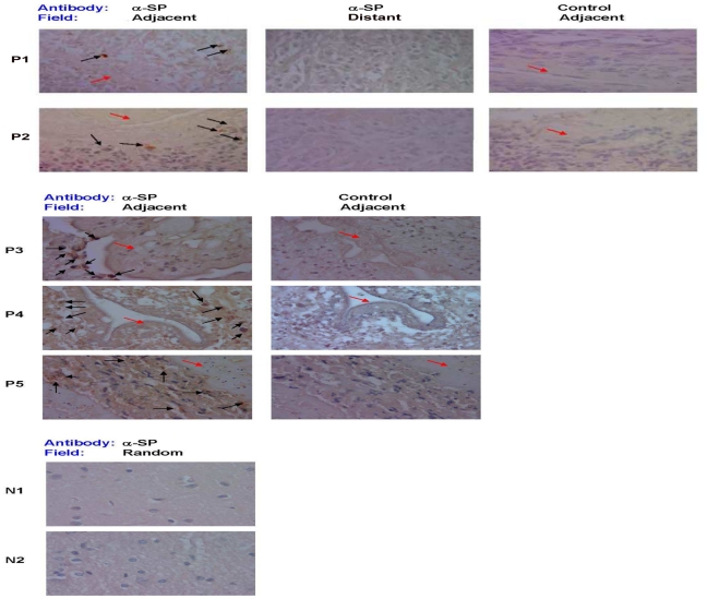
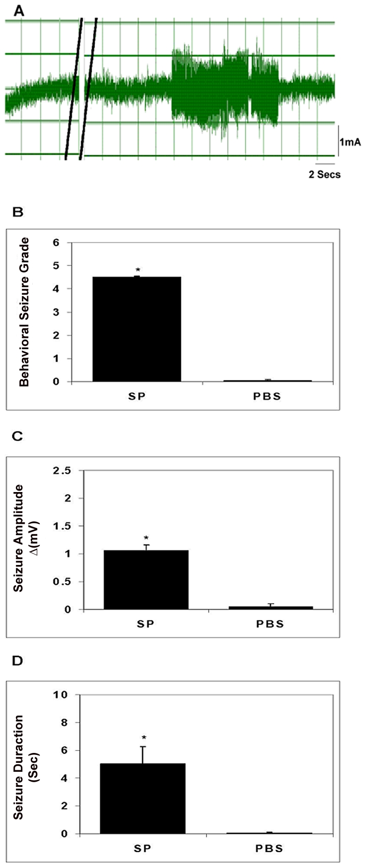
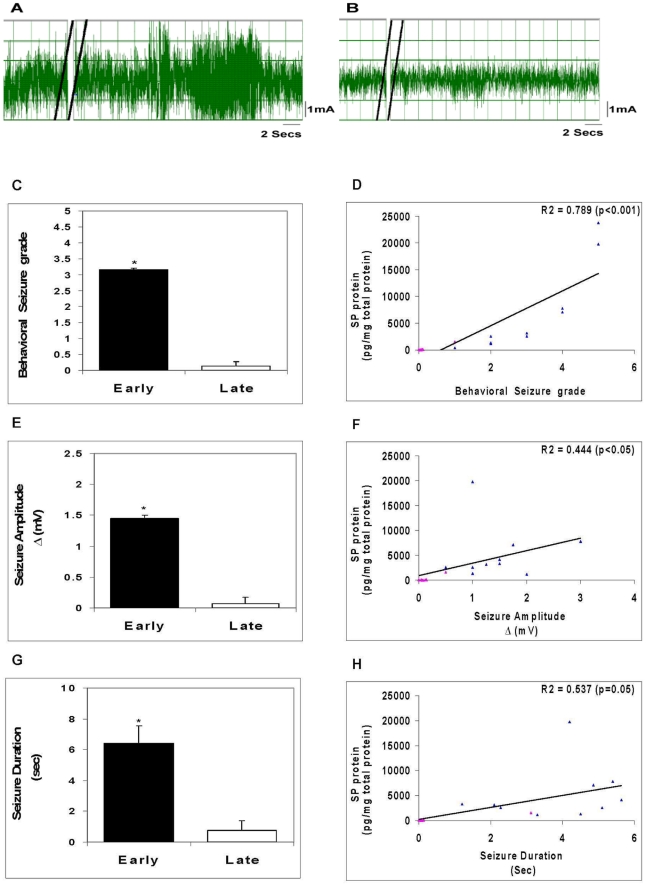
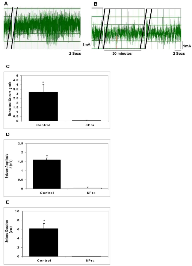
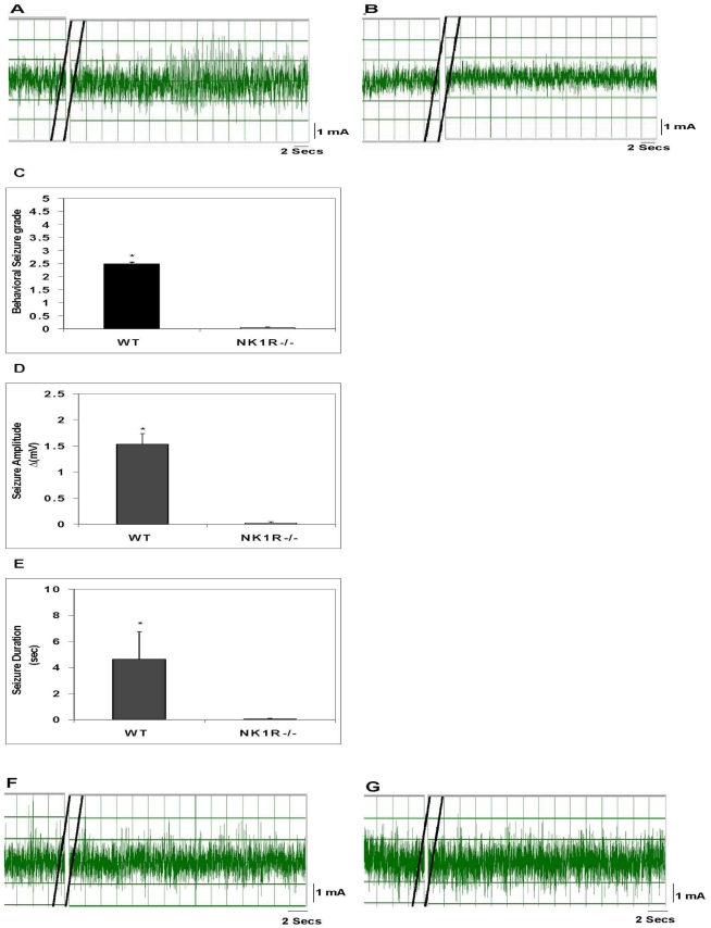
Neurocysticercosis (NCC), a helminth infection of the brain, is a major cause of seizures. The mediators responsible for seizures in NCC are unknown, and their management remains controversial. Substance P (SP) is a neuropeptide produced by neurons, endothelial cells and immunocytes. The current studies examined the hypothesis that SP mediates seizures in NCC. We demonstrated by immunostaining that 5 of 5 brain biopsies from NCC patients contained substance P (SP)-positive (+) cells adjacent to but not distant from degenerating worms; no SP+ cells were detected in uninfected brains. In a rodent model of NCC, seizures were induced after intrahippocampal injection of SP alone or after injection of extracts of cysticercosis granuloma obtained from infected wild type (WT), but not from infected SP precursor-deficient mice. Seizure activity correlated with SP levels within WT granuloma extracts and was prevented by intrahippocampal pre-injection of SP receptor antagonist. Furthermore, extracts of granulomas from WT mice caused seizures when injected into the hippocampus of WT mice, but not when injected into SP receptor (NK1R) deficient mice. These findings indicate that SP causes seizures in NCC, and, suggests that seizures in NCC in humans may be prevented and/or treated with SP-receptor antagonists.


Conflict of interest statement

The authors have declared that no competing interests exist.

Figures

Figure 1
Figure 1. SP immunostaining of brain tissue samples from NCC-infected and uninfected patients.
Photomicrographs of brain biopsy specimens of NCC-infected patients (P1 through P5) and brain autopsy specimens of NCC-uninfected patients (N1 and N2) are shown. Specimen slides were stained with specific polyclonal rabbit anti-SP antibody (α-SP) or control rabbit serum. Fields shown are either adjacent to the parasite (indicated by red arrow) or distant from parasite [when specimen size permitted (P1 and P2)], as indicated. SP+ cells are indicated by black arrows, 1000× magnification. Data shown is representative of one independent experiment.
Figure 2
Figure 2. Seizure activity in rats following intrahippocampal injection of SP.
A representative EEG recording before and after injection of SP into the hippocampus of a rat is shown in panel A; double black lines indicate period of SP injection. The behavioral seizure grade (B), seizure amplitude (C), and seizure duration (D) following intrahippocampal injection of SP or vehicle (PBS) in rats (n = 4–6) are depicted graphically (results shown are mean ± SEM; *, p≤0.001, Mann-Whitney). Results are pooled data from two independent experiments.
Figure 3
Figure 3. Seizure activity in rats following intrahippocampal injection of granuloma extracts.
Representative EEG recordings after injection of an early granuloma extract (A) or a late granuloma extract (B) into the hippocampus of rat are shown; double black lines indicate period of extract injection. The behavioral seizure grade (C), seizure amplitude (E), and seizure duration (G) in rats following intrahippocampal injection of early or late granuloma extracts (n = 7–12) are depicted graphically (*, p≤0.001, Mann-Whitney). Correlation between levels of SP peptide within the granuloma extracts and behavioral seizure grade (D), seizure amplitude (F), and seizure duration (H) along with R2 and p values are shown (blue triangles represent each of the early stage granuloma extracts and pink triangles represent each of the late stage granuloma extracts). Results are pooled data from three independent experiments.
Figure 4
Figure 4. Effect of SP receptor antagonist (SPra) pre-treatment on granuloma extract-induced seizure activity in rats.
Representative EEG recordings in rats after intrahippocampal injection of an early granuloma extract pretreated without (A) or with SPra (B) are shown; double black lines indicate the period of extract injection (A and B) or SP receptor antagonist injection (B only). The behavioral seizure grade (C), seizure amplitude (D), and seizure duration (E) in rats following intrahippocampal injection of early granuloma extracts pre-treated without (Control) or with SPra (SPra; n = 6 each) are depicted graphically (*, p≤0.001, Mann-Whitney). Results are pooled data from two independent experiments.
Figure 5
Figure 5. Effect of deletion of the NK1R gene or the SP gene in mice on granuloma extract-induced seizure activity.
A representative EEG tracing after injection of an early granuloma extract obtained from a wild type mouse into the hippocampus of a wild type mouse (A) or NK1−/− (B) mouse is shown. The behavioral seizure grade (C), seizure amplitude (D), and seizure duration (E) in wild type or NK1−/− mice following intrahippocampal injection of extracts of early granulomas obtained from wild type mice (n = 4 each) are depicted graphically (*, p≤0.001, Mann-Whitney). EEG recordings shown (F and G) are representative of results obtained after intrahippocampal injection into a wild type mouse of an extract from an early granuloma (F) or late granuloma (G) obtained from an SPP−/− mouse. Results are pooled data from two independent experiments.

Similar articles

Cited by

References

    1. Singhi P. Infectious causes of seizures and epilepsy in the developing world. Dev Med Child Neurol. 2011;53:600–609. - PubMed
    1. Shandera WX, Kass JS. Neurocysticercosis: current knowledge and advances. Curr Neurol Neurosci Rep. 2006;6:453–459. - PubMed
    1. Cruz ME, Schantz PM, Cruz I, Espinosa P, Preux PM, et al. Epilepsy and neurocysticercosis in an Andean community. Int J Epidemiol. 1999;28:799–803. - PubMed
    1. Medina MT, Rosas E, Rubio-Donnadieu F, Sotelo J. Neurocysticercosis as the main cause of late-onset epilepsy in Mexico. Arch Intern Med. 1990;150:325–327. - PubMed
    1. White AC., Jr Neurocysticercosis: updates on epidemiology, pathogenesis, diagnosis, and management. Annu Rev Med. 2000;51:187–206. - PubMed

Publication types

MeSH terms