. 2012 Feb;8(2):e1002491.
doi: 10.1371/journal.ppat.1002491. Epub 2012 Feb 9.

The TPR domain in the host Cyp40-like cyclophilin binds to the viral replication protein and inhibits the assembly of the tombusviral replicase

Affiliations

The TPR domain in the host Cyp40-like cyclophilin binds to the viral replication protein and inhibits the assembly of the tombusviral replicase

Jing-Yi Lin et al. PLoS Pathog. 2012 Feb.

Abstract

Replication of plus-stranded RNA viruses is greatly affected by numerous host-coded proteins acting either as susceptibility or resistance factors. Previous genome-wide screens and global proteomics approaches with Tomato bushy stunt tombusvirus (TBSV) in a yeast model host revealed the involvement of cyclophilins, which are a large family of host prolyl isomerases, in TBSV replication. In this paper, we identified those members of the large cyclophilin family that interacted with the viral replication proteins and inhibited TBSV replication. Further characterization of the most effective cyclophilin, the Cyp40-like Cpr7p, revealed that it strongly inhibits many steps during TBSV replication in a cell-free replication assay. These steps include viral RNA recruitment inhibited via binding of Cpr7p to the RNA-binding region of the viral replication protein; the assembly of the viral replicase complex and viral RNA synthesis. Since the TPR (tetratricopeptide repeats) domain, but not the catalytic domain of Cpr7p is needed for the inhibitory effect on TBSV replication, it seems that the chaperone activity of Cpr7p provides the negative regulatory function. We also show that three Cyp40-like proteins from plants can inhibit TBSV replication in vitro and Cpr7p is also effective against Nodamura virus, an insect pathogen. Overall, the current work revealed a role for Cyp40-like proteins and their TPR domains as regulators of RNA virus replication.


Conflict of interest statement

The authors have declared that no competing interests exist.

Figures

Figure 1
Figure 1. Identification of the members of the cyclophilin (immunophilin) family that interact with p33 and inhibit TBSV replication in vitro.
(A) Split ubiquitin MYTH assay was used to test binding between p33 and the shown full-length yeast proteins. The bait p33 was co-expressed with the prey proteins in yeast. SSA1 (HSP70 chaperone), and the empty prey vector (NubG) were used as positive and negative controls, respectively. The interaction of cyclophilins with p33, their localization in cells and human homologs are summarized below the image. (B) Scheme of the CFE-based TBSV replication assay. Purified recombinant p33 and p92pol replication proteins of TBSV and in vitro transcribed TBSV DI-72 (+)repRNA were added to the whole cell extract prepared from various yeast strains with deletion of selected members of the immunophilin genes as shown in panel C. (C) Top panel: Denaturing PAGE analysis of the 32P-labeled TBSV repRNA products obtained in the TBSV replication assay based on various CFEs as shown. Each experiment was repeated three times. Bottom panel: Western blot analysis of various CFEs with anti-Dpm1 antibody, an ER-resident protein, to show similar protein content in CFEs.
Figure 2
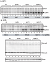
Figure 2. Increased TBSV repRNA accumulation in cpr7Δ yeast.
(A) To launch TBSV repRNA replication, we expressed 6xHis-p33 and 6xHis-p92 from the copper-inducible CUP1 promoter and DI-72(+) repRNA from the galactose-inducible GAL1 promoter in the parental (BY4741) and in cpr6Δ, and cpr7Δ single deletion and cpr6Δ cpr7Δ double deletion yeast strains. The yeast cells were cultured for 40 hours at either 29°C (panel A) or 23°C (panel B) on 2% galactose SC minimal media. Northern blot analysis was used to detect DI-72(+) repRNA accumulation. The accumulation level of DI-72(+) repRNA was normalized based on 18S rRNA. Bottom panels: Western blot analysis of the accumulation level of 6xHis-tagged p33 and 6xHis-tagged p92 proteins using anti-His antibody. Asterisk marks the SDS-resistant p33 homodimer. The Western blot for PGK host protein at the bottom shows the loading control.
Figure 3
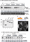
Figure 3. Cell-free TBSV replication assay supports an inhibitory role for Cpr7p and an Arabidopsis Cyp40 homolog.
(A) Top panel: Denaturing PAGE analysis of the 32P-labeled TBSV repRNA products obtained in the CFE-based assay programmed with in vitro transcribed TBSV DI-72 (+)repRNA and purified recombinant p33 and p92pol replication proteins of TBSV. Purified recombinant GST-tagged Cpr7p, the Cyp domain or the TPR domain (0.4, 0.8 and 1.6 µg), or GST were added to CFE prepared from BY4741 yeast strain. Each experiment was repeated three times. Middle panel: Ethidium-bromide stained PAGE gel from the top panel to show the sample loading and the lack of RNase activity in the CFE-based assay. Bottom panel: SDS-PAGE analysis of the purified recombinant proteins used in the above CFE-based assay. (B) Split ubiquitin assay was used to demonstrate binding between p33 and the TPR and the catalytic Cyp domains of Cpr7p. The domain structure and size of Cpr7p is shown on the top. (C) Denaturing PAGE analysis of the 32P-labeled TBSV repRNA products obtained in the CFE-based TBSV replication assay containing purified recombinant Arabidopsis Cyp40 homolog At5g48570 and its TPR domain (0.4, 0.8 and 1.6 µg).
Figure 4
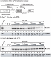
Figure 4. Step-wise CFE-based TBSV replication assay supports an inhibitory role for Cpr7p in the assembly of the TBSV VRC.
(A) Scheme of the CFE-based TBSV replicase assembly and replication assays. Purified recombinant p33 and p92pol replication proteins of TBSV and in vitro transcribed TBSV DI-72 (+)repRNA were added to CFE prepared from BY4741 yeast strain in step 1. The assay either contained or lacked the purified recombinant Cpr7p, its derivatives (0.4, 0.8 and 1.6 µg), or GST during step 1. Note that the assay was performed in the presence of ATP/GTP to facilitate TBSV VRC assembly, but prevent RNA synthesis in step 1. After step 1, centrifugation was used to collect the membrane fraction of the CFE, and after washing the membranes, step 2 was performed in the presence of ATP/CTP/GTP and 32P-UTP to allow TBSV RNA replication. In the samples presented on the right side of the panel, the recombinant Cpr7p or derivatives were added at the beginning of step 2. (B) Denaturing PAGE analysis of the 32P-labeled TBSV repRNA products obtained in the CFE-based assays when Cpr7p or derivatives were added at the 1st step. See further details in Figure 1. (C) Denaturing PAGE analysis of the 32P-labeled TBSV repRNA products obtained in the CFE-based assays when Cpr7p or derivatives were added at the 2nd step. Three repeats of each experiment were performed.
Figure 5
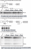
Figure 5. Cpr7p affects multiple steps during TBSV RNA replication in vitro.
(A) Scheme of the viral RNA recruitment assay based on CFE. The 32P-labeled TBSV (+)repRNA template was added together with p33/p92 and Cpr7p or its derivatives to CFE prepared from BY4741 yeast. The membrane-association of 32P-labeled TBSV (+)repRNA template is measured using denaturing PAGE gels (Panel B). Note that 32P-labeled TBSV (+)repRNA template can inefficiently associate with the membrane even in the absence of p33/p92 (lane 2), likely due to nonspecific binding to an RNA-binding host protein in the membrane. (C) Scheme of the CFE-based TBSV replicase assembly assay. Note that the original template RNA is removed during replicase solubilization/purification. Therefore, an added (−)repRNA is tested for replicase activity for each replicase prep. (D) Complementary RNA synthesis that results in 32P-labeled TBSV (+)repRNA template is measured using denaturing PAGE gels. (E) Detection of single- and double-stranded RNA products produced in the CFE-based TBSV replication assay. The ratio of ssRNA and dsRNA in the samples are shown. Note that the dsRNA product represents the annealed 32P-labeled (−)RNA and the (+)RNA, while the ssRNA products represents the newly made 32P-labeled (+)RNA products.
Figure 6
Figure 6. Cpr7p inhibits complementary RNA synthesis by the affinity-purified tombusvirus replicase.
(A) Representative denaturing gel of 32P-labeled RNA products synthesized by the purified tombusvirus replicase in vitro in the presence of 0.8, 1.2 and 1.6 µg of purified recombinant Cpr7p or GST. The in vitro assays were programmed with DI-72 (−)repRNA, and they also contained ATP/CTP/GTP and 32P-UTP. All the components were added at the same time. The level of complementary RNA synthesis producing “FL” (the full-length product, made via initiation from the 3′-terminal promoter, also called “ti” product) is shown as % of FL product in the control sample. Note that this replicase preparation also synthesizes internal initiation products (“ii”) and 3′-terminal extension products (“3′TEX”). Each experiment was repeated three times. (B) The in vitro replicase experiment is similar to that in Panel A, except GST-Cpr7 or GST was pre-incubated for 10 min with the replicase to facilitate binding of Cpr7p and the VRC prior to RNA synthesis.
Figure 7
Figure 7. Binding of Cpr7p to TBSV p33 protein derivatives in vitro.
(A) Schematic representation of viral p33 and its derivatives used in the binding assay. The various domains include: TMD, transmembrane domain; RPR, arginine-proline-rich RNA binding domain; P; phosphorylated serine and threonine; S1 and S2 subdomains involved in p33:p33/p92 interaction. (B) Affinity binding (pull-down) assay to detect interaction between FLAG-Cpr7p and the MBP-tagged viral p33 protein derivatives. The MBP-tagged viral proteins and MBP produced in E. coli were immobilized on amylose-affinity columns. Then, FLAG-Cpr7p expressed in BY4741 yeast was passed through the amylose-affinity columns with immobilized MBP-tagged proteins. The affinity-bound proteins were eluted with maltose from the columns. The eluted proteins were analyzed by Western blotting with anti-FLAG antibody to detect the amount of FLAG-Cpr7p specifically bound to MBP-tagged viral proteins. (C) Pull-down assay to detect interaction between FLAG-Cpr7p, FLAG-Cpr6p and the MBP-tagged p33CΔRPR, a mutated viral p33 protein lacking the RPR motif that binds to the viral RNA. See further details in Panel B.
Figure 8
Figure 8. Increased NoV and FHV RNA accumulation in cpr7Δ yeast.
Split ubiquitin MYTH assay was used to test binding between the yeast cyclophilins and NoV protein A (panel A), TMV 130K (panel B) and TCV p28 (panel C) replication proteins. The bait proteins were co-expressed with the prey cyclophilin proteins in yeast. The empty prey vector (NubG) was used as a negative control. To launch NoV (panel D) or FHV RNA1 (panel E) replication, we expressed NoV RNA1 from the copper-inducible CUP1 promoter and FHV RNA1 and DI634 from the CUP1 promoter in the parental (BY4741) and in cpr6Δ, and cpr7Δ single deletion and cpr6Δ cpr7Δ double deletion yeast strains. The yeast cells were cultured for 48 hours at 29°C in 3 ml SC-H with 2% glucose media containing 50 mM CuSO4 (for NoV) and for 24 hours at 29°C in 3 ml SC-H with 2% galactose (for FHV). Northern blot analysis was used to detect RNA1/RNA3 accumulation for NoV and FHV. The accumulation level of NoV and FHV RNAs was normalized based on 18S rRNA. Each experiment was repeated.
Figure 9
Figure 9. Models for the role Cpr7p in tombusvirus replication.
Model 1: Cpr7p has been shown to inhibit the recruitment of the viral RNA into replication. Model 2: The assembly of the tombusviral VRC requires interactions among the viral replication proteins, selected host proteins, such as Hsp70, GAPDH or eEF1A or membrane lipids. Cpr7p might inhibit any of these interactions, resulting in reduced TBSV RNA replication. In addition, Cpr7p binds to the RPR RNA binding motif in p33 and likely inhibiting p33: viral (+)RNA interaction that is needed for VRC assembly. Model 3: Due to the chaperone activity of the TPR domain, binding of Cpr7p to p92 might inhibit the RdRp function of p92, thus blocking RNA synthesis. See further details in the text.

Similar articles

Cited by

References

    1. Nagy PD. Yeast as a model host to explore plant virus-host interactions. Annu Rev Phytopathol. 2008;46:217–242. - PubMed
    1. Nagy PD, Pogany J. Multiple roles of viral replication proteins in plant RNA virus replication. Methods Mol Biol. 2008;451:55–68. - PubMed
    1. Nagy PD, Wang RY, Pogany J, Hafren A, Makinen K. Emerging picture of host chaperone and cyclophilin roles in RNA virus replication. Virology. 2011;411:374–382. - PubMed
    1. Li Z, Nagy PD. Diverse roles of host RNA binding proteins in RNA virus replication. RNA Biol. 2011;8:305–315. - PMC - PubMed
    1. den Boon JA, Diaz A, Ahlquist P. Cytoplasmic viral replication complexes. Cell Host Microbe. 2010;8:77–85. - PMC - PubMed

Publication types

MeSH terms