. 2012 Feb;8(2):e1002521.
doi: 10.1371/journal.pgen.1002521. Epub 2012 Feb 9.

Genetic and functional analyses of SHANK2 mutations suggest a multiple hit model of autism spectrum disorders

Affiliations

Genetic and functional analyses of SHANK2 mutations suggest a multiple hit model of autism spectrum disorders

Claire S Leblond et al. PLoS Genet. 2012 Feb.

Abstract

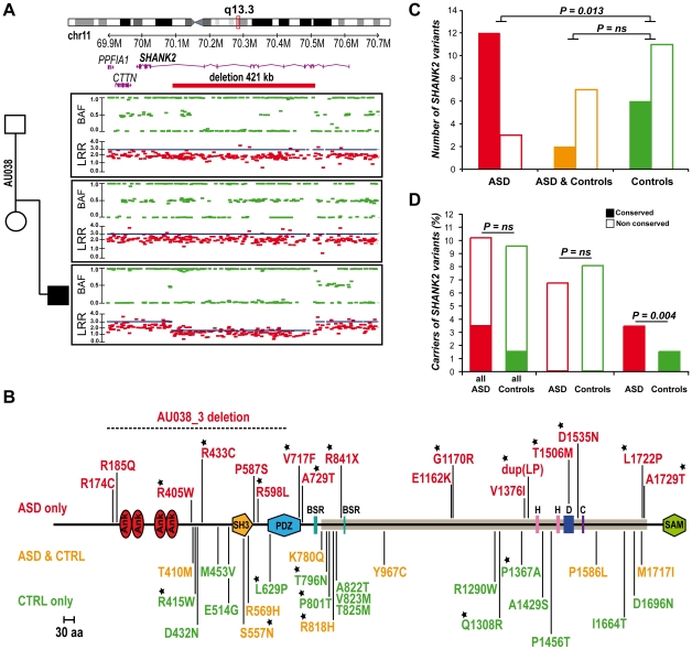
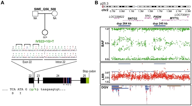
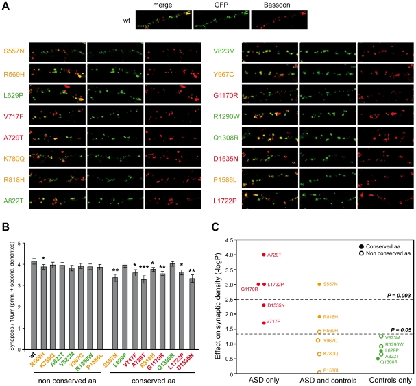
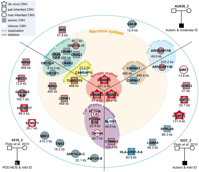
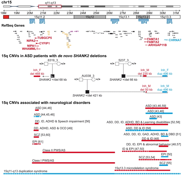
Autism spectrum disorders (ASD) are a heterogeneous group of neurodevelopmental disorders with a complex inheritance pattern. While many rare variants in synaptic proteins have been identified in patients with ASD, little is known about their effects at the synapse and their interactions with other genetic variations. Here, following the discovery of two de novo SHANK2 deletions by the Autism Genome Project, we identified a novel 421 kb de novo SHANK2 deletion in a patient with autism. We then sequenced SHANK2 in 455 patients with ASD and 431 controls and integrated these results with those reported by Berkel et al. 2010 (n = 396 patients and n = 659 controls). We observed a significant enrichment of variants affecting conserved amino acids in 29 of 851 (3.4%) patients and in 16 of 1,090 (1.5%) controls (P = 0.004, OR = 2.37, 95% CI = 1.23-4.70). In neuronal cell cultures, the variants identified in patients were associated with a reduced synaptic density at dendrites compared to the variants only detected in controls (P = 0.0013). Interestingly, the three patients with de novo SHANK2 deletions also carried inherited CNVs at 15q11-q13 previously associated with neuropsychiatric disorders. In two cases, the nicotinic receptor CHRNA7 was duplicated and in one case the synaptic translation repressor CYFIP1 was deleted. These results strengthen the role of synaptic gene dysfunction in ASD but also highlight the presence of putative modifier genes, which is in keeping with the "multiple hit model" for ASD. A better knowledge of these genetic interactions will be necessary to understand the complex inheritance pattern of ASD.


Conflict of interest statement

The authors have declared that no competing interests exist.

Figures

Figure 1
Figure 1. Genomic structure, isoforms, and expression of human SHANK2.
A. Genomic structure of the human SHANK2 gene. Transcription of SHANK2 produces four main mRNA from three distinct promoters: SHANK2E (AB208025), ProSAP1A (AB208026), ProSAP1 (AB208027) and AF141901. There are three translation starts: in exon 2 for SHANK2E, in exon1b for ProSAP1A, and in exon1c for ProSAP1 and AF141901; and two independent stop codons: in exon 22b for AF141901 and in exon 25 for SHANK2E, ProSAP1A and ProSAP1. Conserved domains of protein interaction or protein binding site are represented in color: ANK (red), SH3 (orange), PDZ (blue) and SAM (green), H (pink), D, (dark blue) and C (purple). Black stars identify the alternative spliced exons (‘brain-specific exons’ in turquoise: 19, 20 and 23). B. RT-PCRs of SHANK2 isoforms on RNA from different human control tissues (Clontech), and different brain regions of four controls (2 males and 2 females). The amplified regions specific to each isoform of SHANK2 are indicated by gray boxes. C. Alternative splicing of human SHANK2; exons 19, 20 and 23 are specific to the brain. ANK, ankyrin; SH3, Src homology 3; PDZ, PSD95/DLG/ZO1; SAM, sterile alpha motif; He, heart; Li, liver; B, brain; SM, skeletal muscle; Pl, placenta; K, kidney; Lu, lung; Pa, pancreas; FC, frontal cortex; Hi, hippocampus; TC, temporal cortex; T, thalamus; OC, occipital cortex; Ce, cerebellum; Cx, whole cortex; BLCL, B lymphoblastoid cell lines; GAPDH, glyceraldehyde 3-phosphate dehydrogenase; BSR, brain specific region; H, homer binding site; D, dynamin binding site; C, cortactin binding site. The ages of the two males and the two females studied were 74, 42, 55, and 36 years with a post-mortem interval of 10, 21, 24, and 2 h, respectively.
Figure 2
Figure 2. SHANK2 mutations in patients with ASD.
A. A heterozygous deletion of SHANK2 was identified with the Illumina Human 1M-Duo SNP array in a patient with autism (AU038_3). The deletion spans 421 kb on chromosome 11q13.3, covers twelve exons of the human SHANK2 and is not present in the parents. Each dot shows Log R Ratio (LRR; in red) and B allele frequency (BAF; in green). QuantiSNP score is represented with a blue line and indicates the deletion size. B. Location of the CNV and sequence variants (from this study and Berkel et al. 2010) along the SHANK2 protein: in red the variations specific to ASD, in orange the variations shared by ASD and controls and in green the variations specific to controls . The breakpoints of the SHANK2 deletion in AU038_3 are represented with a dotted line on the protein. Stars indicate the variants affecting conserved amino acids. C. A total of 40 variants were identified and variants affecting conserved amino acids in other SHANK proteins are enriched in patients with ASD (nconserved = 12 and nnon-conserved = 3) compared with controls (nconserved = 6 and nnon-conserved = 11) (Fisher's exact test 1-sided, P = 0.013, OR = 6.83, 95% IC = 1.19–53.40). D. The percentage of carriers of SHANK2 variants in patients with ASD and Controls. Variants affecting a conserved amino acid among the SHANK proteins are enriched in patients with ASD (nconserved = 29 and nnon-conserved = 822) compared with controls (nconserved = 16 and nnon-conserved = 1074) (Fisher's exact test 1-sided, P = 0.004, OR = 2.37, 95% CI = 1.23–4.70). Open squares and filled squares represent the non-conserved and conserved amino acids, respectively. ANK, Ankyrin repeat domain; SH3, Src homology 3 domain; PDZ, postsynaptic density 95/Discs large/zona occludens-1 homology domain; SAM, sterile alpha motif domain; BSR, brain specific region; H, homer binding site; D, dynamin binding site; C, cortactin binding site. The proline-rich region is represented as a horizontal gray line.
Figure 3
Figure 3. Genetic alterations identified in the control subject SWE_Q56_508.
A. SHANK2 splice mutation (IVS22+1G>T) detected in a Swedish female control, SWE_Q56_508. The mutation altered the donor splicing site of exon 22 and led to a premature stop in all SHANK2 isoforms except for the AF1411901 isoform, where it altered the protein sequence (G263V). B. CNVs in the same individual altering LOC339822, SNTG2, PXDN and MYT1L. The two close duplications span 264 kb and 245 kb on chromosome 2 and altered LOC339822 and SNTG2, and PXDN and MYT1L, respectively. Dots show the B allele frequency (BAF; in green), Log R ratio (LRR; in red), and QuantiSNP score (in blue). Lower panel: all CNVs listed in the Database of Genomic Variants (DGV) are represented: loss (in red), gain (in blue), gain or loss (in brown). H, homer binding site; D, dynamin binding site; C, cortactin binding site.
Figure 4
Figure 4. Characterization of the functional impact of SHANK2 mutations in cultured neuronal cells.
A. The colocalization of ProSAP1A/SHANK2-EGFP (postsynaptic marker) and Bassoon (presynaptic marker) indicated that the mutations did not disturb the formation of SHANK2 clusters at excitatory synapses along the dendrites. B. The quantification of synapse density was performed on 20 transfected hippocampal neurons per construct from at least three independent experiments. The majority of the ProSAP1A variants affecting a conserved amino acid among SHANK proteins reduced significantly the synaptic density compared with the variants that affect amino acid non conserved among SHANK proteins (Mann-Whitney U-test: nWT = 20, nmut = 20; US557N = 82.5, pS557N = 0.001; UR569H = 124, pR569H = 0.04; UL629P = 149, pL629P = 0.17; UV717F = 114, pV717F = 0.02; UA729T = 73, pA729T = 0.000; UK780Q = 154, pK780Q = 0.221; UR818H = 108, pR818H = 0.012; UA822T = 154.5, pA822T = 0.224; UV823M = 129, pV823M = 0.056; UY967C = 134, pY967C = 0.076; UG1170R = 78, pG1170R = 0.001; UR1290W = 142, pR1290W = 0.121; UQ1308R = 162, pQ1308R = 0.314; UD1535N = 97, pD1535N = 0.005; UP1586L = 137, pP1586L = 0.910; UL1722P = 79, pL1722P = 0.001, *p<0.05, **p<0.01, ***p<0.001). C. Effect of the variants on synaptic density. The y-axis represents −log P compared to WT (P obtained with Mann-Whitney test). After Bonferroni correction for 16 tests, only P values<0.003 were considered as significant. Variants represented in red were specific to ASD, in orange were shared by ASD and controls, and in green were specific to the controls. Open circles and filled circles represent non conserved and conserved amino acids, respectively. Prim, primary; second, secondary.
Figure 5
Figure 5. Characterization of CNVs in three patients carrying a de novo deletion of SHANK2.
Paternally or maternally inherited CNVs are indicated by squares and circles, respectively. De novo CNVs are indicated by stars. Deletions and duplications are indicated in red and blue, respectively. CNVs hitting exons or only introns are filled with grey and white, respectively. Squares and circles within star represent de novo CNV of paternal or maternal origin; circles within squares represent CNV inherited by father or mother. ABCC6, ATP-binding cassette, sub-family C, member 6 pseudogene 2; ADAM, ADAM metallopeptidase; AMY1, amylase (salivary); AMY2A, amylase (pancreatic); ARHGAP11B, Rho GTPase activating protein 11B; CAMSAP1L1, calmodulin regulated spectrin-associated protein 1-like 1; CHRNA7, cholinergic receptor, nicotinic, alpha 7; CNTN4, contactin 4; CTNNA3, catenin (cadherin-associated protein), alpha 3; CYFIP1, cytoplasmic FMR1 interacting protein 1; DUSP22, dual specificity phosphatase 22; GALM, galactose mutarotase; GCNT2, glucosaminyl (N-acetyl) transferase 2; GOLGA, golgi autoantigen, golgin subfamily a; GSTT1, glutathione S-transferase theta 1; HLA-DRB, major histocompatibility complex, class II, DR beta; LAMA4, laminin, alpha 4; NIPA, non imprinted in Prader-Willi/Angelman syndrome; NLGN1, neuroligin 1; NME7, non-metastatic cells 7; OR, olfactory receptor; PCDHA, protocadherin alpha; RFPL4B, ret finger protein-like 4B; RHD, Rh blood group, D antigen; SFMBT1, Scm-like with four mbt domains 1; SHANK2, SH3 and multiple ankyrin repeat domains 2; SMC2, structural maintenance of chromosomes 2; TNS3, tensin 3; TUBGCP5, tubulin, gamma complex associated protein 5; UGT2B17, UDP glucuronosyltransferase 2 family, polypeptide B17.
Figure 6
Figure 6. Inherited 15q11–q13 CNVs identified in three ASD patients carrier of a de novo SHANK2 deletion.
Deletions (del) and duplications (dup) are indicated in red and blue, respectively. Paternally and maternally imprinted genes are indicated in yellow and pink, respectively. Genes altered by the CNVs are indicated in blue or red. The bottom part of the figure indicates the location of the deletions/duplications previously associated with neuropsychiatric disorders –. BP, breakpoint; Inh_M, inherited by mother; Inh_F, inherited by father; AS, Angelman syndrome; ASD, Autism spectrum disorders; ADHD, attention deficit-hyperactivity disorder; BP, bipolar disorder; DD: developmental delay; DBD, disruptive behavior disorder; EPI, epilepsy; GAD, generalized anxiety disorder; OCD, obsessive-compulsive disorder; ID, intellectual disability; PWS, Prader-Willi syndrome; SCZ, schizophrenia.

Similar articles

Cited by

References

    1. American Psychiatric Association. Diagnostic and Statistical Manual of Mental Disorders, 4th Ed. American Psychiatric Press, Washington D.C; 1994.
    1. Fernell E, Gillberg C. Autism spectrum disorder diagnoses in Stockholm preschoolers. Res Dev Disabil. 2010;31:680–685. - PubMed
    1. Freitag CM. The genetics of autistic disorders and its clinical relevance: a review of the literature. Mol Psychiatry. 2007;12:2–22. - PubMed
    1. Hallmayer J, Cleveland S, Torres A, Phillips J, Cohen B, et al. Genetic Heritability and Shared Environmental Factors Among Twin Pairs With Autism. Arch Gen Psychiatry 2011 - PMC - PubMed
    1. Bourgeron T. A synaptic trek to autism. Curr Opin Neurobiol. 2009;19:231–234. - PubMed

Publication types

MeSH terms