. 2012 Feb 6:6:13.
doi: 10.3389/fnins.2012.00013. eCollection 2012.

Fine-Tuning Oligodendrocyte Development by microRNAs

Affiliations

Fine-Tuning Oligodendrocyte Development by microRNAs

Olga Barca-Mayo et al. Front Neurosci. .

Abstract

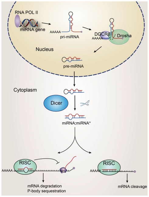
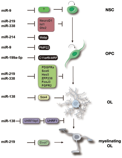
Myelination of axons by oligodendrocytes in the central nervous system is essential for normal neuronal functions. The failure of remyelination due to injury or pathological insults results in devastating demyelinating diseases. Oligodendrocytes originate in restricted regions of the embryonic ventral neural tube. After migration to populate all areas of the brain and spinal cord, oligodendrocyte precursors undergo a temporally well-defined series of molecular and structural changes, ultimately culminating in the cessation of proliferation, and the elaboration of a highly complex myelin sheath. The emergence of microRNAs (miRNAs) as potent regulators of gene expression at the posttranscriptional level has broad implications in all facets of cell biology. Recent studies have demonstrated a critical role of miRNAs in oligodendrocyte development, including cell proliferation, differentiation, and myelin formation. In this review, we will highlight and discuss the recent understanding of functional links of miRNAs to regulatory networks for central myelination, as well as perspectives on the role of miRNAs in demyelinating diseases.

Keywords: feed-back regulation; miRNAs; myelination; neural cell fate; oligodendrocyte; transcriptional control.


Figures

Figure 1
Figure 1
Biosynthesis of miRNAs. The miRNA gene is transcribed by RNA polymerase II as pri-miRNAs which are then processed by Drosha to produce long miRNA precursor (pre-miRNA). The pre-miRNAs are transported to the cytoplasm where they are further cleaved by the endoribonuclease Dicer to mature ∼22 nt long miRNA–miRNA *duplex. In this process Dicer interacts with the TRBP, PACT, and Argonaute proteins, which form the RNA-induced silencing complex (RISC). Within this complex, one strand of the miRNA duplex is removed and the single stranded miRNA, complementary to the target mRNA, remains in the complex and becomes functional. Seven to eight base pair “seed” sequence in 5′ miRNAs is partially complementary to the 3 UTR of mRNA targets, and induces posttranscriptional silencing through mechanisms such as mRNA destabilization and translational repression.
Figure 2
Figure 2
miRNAs function at multiple stages to control oligodendrocyte differentiation and myelination. Specific miRNAs involved in regulating the neural precursor to OPC transition, OPC differentiation, OL maturation and myelination as well as the documented and predicted targets of those miRNAs are shown. miR-9, miR-214, and miR-199a-5p are highly expressed in OPCs and prevent premature differentiation by blocking expression of myelin components including Mobp, PMP22 and maturation promoting factors such as MRF/C11orf9. miR-138, -219 and -338 are more highly expressed in OLs and promote OL differentiation by inhibiting expression of differentiation inhibitors (e.g., Hes5, Sox6, and Sox4) and proliferation signaling molecules such as PDGFRα and FGFR, as well as neuronal differentiation factors (e.g., Zfp238 and FoxJ3). miR-219 may also regulate OL terminal maturation and myelin maintenance by targeting fatty acid elongases such as Elvol7. The expression pattern of miRNAs is likely correlated to their functions at different stages of OL lineage progression.

Similar articles

Cited by

References

    1. Asirvatham A. J., Gregorie C. J., Hu Z., Magner W. J., Tomasi T. B. (2008). MicroRNA targets in immune genes and the Dicer/Argonaute and ARE machinery components. Mol. Immunol. 45, 1995–200610.1016/j.molimm.2007.10.035 - DOI - PMC - PubMed
    1. Babiarz J. E., Ruby J. G., Wang Y., Bartel D. P., Blelloch R. (2008). Mouse ES cells express endogenous shRNAs, siRNAs, and other microprocessor-independent, Dicer-dependent small RNAs. Genes Dev. 22, 2773–278510.1101/gad.1705308 - DOI - PMC - PubMed
    1. Bartel D. P. (2009). MicroRNAs: target recognition and regulatory functions. Cell 136, 215–23310.1016/j.cell.2009.01.002 - DOI - PMC - PubMed
    1. Barres B. A., Raff M. C. (1994). Control of oligodendrocyte number in the developing rat optic nerve. Neuron 12, 935–94210.1016/0896-6273(94)90305-0 - DOI - PubMed
    1. Brett J. O., Renault V. M., Rafalski V. A., Webb A. E., Brunet A. (2011). The microRNA cluster miR-106b∼25 regulates adult neural stem/progenitor cell proliferation and neuronal differentiation. Aging (Albany N. Y.) 3, 108–124 - PMC - PubMed

LinkOut - more resources