. 2012;7(2):e29948.
doi: 10.1371/journal.pone.0029948. Epub 2012 Feb 8.

Structural and functional insights into endoglin ligand recognition and binding

Affiliations

Structural and functional insights into endoglin ligand recognition and binding

Aaron Alt et al. PLoS One. 2012.

Abstract

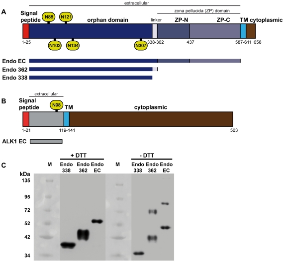
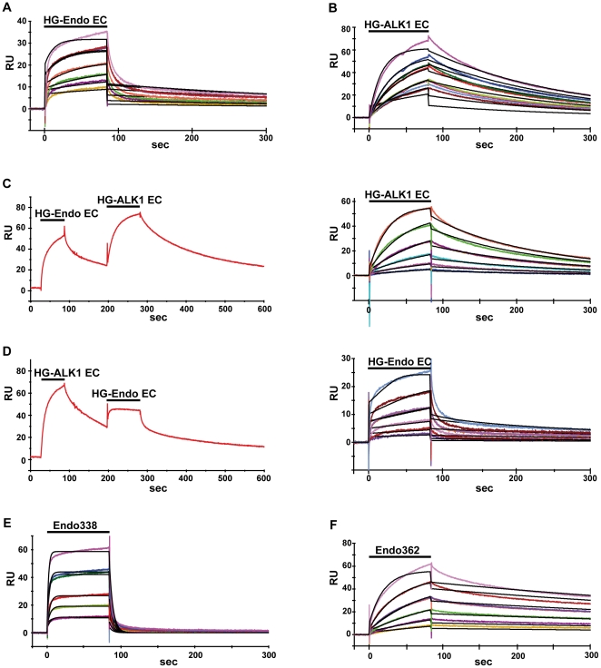
Endoglin, a type I membrane glycoprotein expressed as a disulfide-linked homodimer on human vascular endothelial cells, is a component of the transforming growth factor (TGF)-β receptor complex and is implicated in a dominant vascular dysplasia known as hereditary hemorrhagic telangiectasia as well as in preeclampsia. It interacts with the type I TGF-β signaling receptor activin receptor-like kinase (ALK)1 and modulates cellular responses to Bone Morphogenetic Protein (BMP)-9 and BMP-10. Structurally, besides carrying a zona pellucida (ZP) domain, endoglin contains at its N-terminal extracellular region a domain of unknown function and without homology to any other known protein, therefore called the orphan domain (OD). In this study, we have determined the recognition and binding ability of full length ALK1, endoglin and constructs encompassing the OD to BMP-9 using combined methods, consisting of surface plasmon resonance and cellular assays. ALK1 and endoglin ectodomains bind, independently of their glycosylation state and without cooperativity, to different sites of BMP-9. The OD comprising residues 22 to 337 was identified among the present constructs as the minimal active endoglin domain needed for partner recognition. These studies also pinpointed to Cys350 as being responsible for the dimerization of endoglin. In contrast to the complete endoglin ectodomain, the OD is a monomer and its small angle X-ray scattering characterization revealed a compact conformation in solution into which a de novo model was fitted.


Conflict of interest statement

Competing Interests: The authors have declared that no competing interests exist.

Figures

Figure 1
Figure 1. Schematic domain organization of human endoglin and ALK1 and western blots of endoglin domains.
Bar diagram of (A) endoglin and (B) ALK1 with the domains indicated and highlighted in different styles. TM, transmembrane region, ZP, zona pellucida. The putative Asn glycosylation sites Asn88, Asn102, Asn121, Asn134 and Asn306 of endoglin and Asn98 of ALK1 are labelled within green ovals. The constructs used in this study and the domains encompassed by these are shown below the bar diagram of the respective full length protein. (C) Western blots of endoglin constructs. Endo338, Endo362 and LG-EndoEC were analyzed by 10% SDS polyacrylamide electrophoresis gel followed by western blotting with an anti-His6 antibody. Samples reduced with dithithreitol (DTT) were incubated for 1 h with 10 mM of this reagent at 65°C. All samples (0.5 µg of protein) were then denaturated by boiling at 95°C for 5 minutes prior to charging onto the gel. The molecular weight markers (M) are indicated at the left of the samples. For both Endo362 and LG-EndoEC, dimeric species are visible, while Endo338 was only observed in monomeric form.
Figure 2
Figure 2. Functional interactions between endoglin, ALK1 and BMP-9.
The interactions of (A) EndoEC, (B) ALK1EC, (E) Endo338 and (F) Endo362 with BMP-9 were investigated by SPR. While HG-EndoEC and Endo362 dissociated slowly, Endo338 dissociated much faster, indicating a rigid body type of binding as opposed to an induced fit mechanism. For affinity measurements, the indicated recombinant proteins were injected at six concentrations ranging from 12.5 to 400 nM over BMP-9 (which was immobilized on a CM5 sensor chip by amine coupling) to generate sensorgrams (colored curves). When testing competition between HG-EndoEC and HG-ALK1EC (C and D) the chip was first pre-equilibrated with 750 mM of either HG-EndoEC (C, left) or HG-ALK1EC (D, left) before injecting the various concentration of the second ligand, showing the curve for the highest concentrations of the 2nd ligand. Both HG-ALK1EC (C, right) and HG-EndoEC (D, right) yielded, after subtracting the background, similar results to those in runs in which no first ligand was preequilibrated before injecting the second ligand (D right vs. E; C right vs. B). This leads to the conclusion that endoglin and ALK1 bind independently to different sites on BMP-9. The kinetic parameters for the interaction were determined by global fitting (curves in black) of the 1∶1 Langmuir binding model to these data, providing values for the association (ka) and dissociation (kd) rate constants and the dissociation affinity constant (KD).
Figure 3
Figure 3. Functional analysis of recombinant endoglin and ALK1 proteins.
ID1 expression in BMP-9 stimulated HMEC-1 cells after 36 hours in the presence or absence of LG-ALK1EC, Endoglin338 or LG-EndoEC. The ALK1 and endoglin ectodomains hijack BMP-9 and therefore signaling is diminished. Endo338 only partially inhibit signaling, due to a less stable complex formed between the orphan domain and the cytokine. (*) Statistically significant (p<0.01) difference compared to unstimulated control cells (Ctl). (#) Statistically significant (p<0.01) difference compared to ID1 expression of BMP-9 stimulated cells.
Figure 4
Figure 4. SAXS data analysis and rigid body modeling.
(A) The experimental SAXS profile of recombinant Endo338, measured at the ID14-3 BioSAXS beamline. The Kratky plot (inserted), displays a gaussian behavior, which indicates the well folded nature of the construct. (B) The distance distribution function, p(r) obtained from GNOM with a Dmax of 9.4 nm also suggests a compact structure. (C) The orphan domain model obtained from I-TASSER together with 5 Man5NAG2 glycans, restricted by their attachment sites was fitted by rigid body modeling with SASREF, yielding a fit of the model (red line) against the experimental Endo338 scattering profile (blue crosses) of χ2 = 1.1. The input models used, are depicted above the fit curve. (D) The resulting model is displayed as a cartoon in three orthogonal views, with the attached sugars depicted as balls, fitted into the molecular envelope from DAMMIF.

References

    1. Bernabeu C, Lopez-Novoa JM, Quintanilla M. The emerging role of TGF-beta superfamily coreceptors in cancer. Biochim Biophys Acta. 2009;1792:954–973. - PubMed
    1. Lebrin F, Mummery CL. Endoglin-mediated vascular remodeling: mechanisms underlying hereditary hemorrhagic telangiectasia. Trends Cardiovasc Med. 2008;18:25–32. - PubMed
    1. Lopez-Novoa JM, Bernabeu C. The physiological role of endoglin in the cardiovascular system. Am J Physiol Heart Circ Physiol. 2010;299:H959–974. - PubMed
    1. McAllister KA, Grogg KM, Johnson DW, Gallione CJ, Baldwin MA, et al. Endoglin, a TGF-beta binding protein of endothelial cells, is the gene for hereditary haemorrhagic telangiectasia type 1. Nat Genet. 1994;8:345–351. - PubMed
    1. Bernabeu C, Blanco FJ, Langa C, Garrido-Martin EM, Botella LM. Involvement of the TGF-beta superfamily signalling pathway in hereditary haemorrhagic telangiectasia. J Appl Biomed. 2010;8:169–171.

Publication types