

Creatine protects against excitoxicity in an in vitro model of neurodegeneration
- PMID: 22347384
- PMCID: PMC3275587
- DOI: 10.1371/journal.pone.0030554
Creatine protects against excitoxicity in an in vitro model of neurodegeneration
Abstract
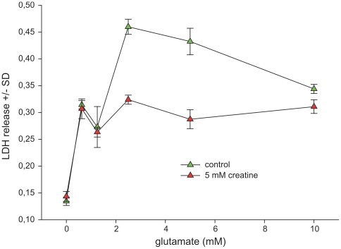
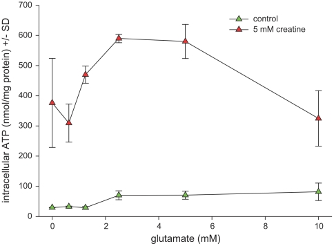
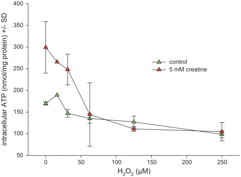
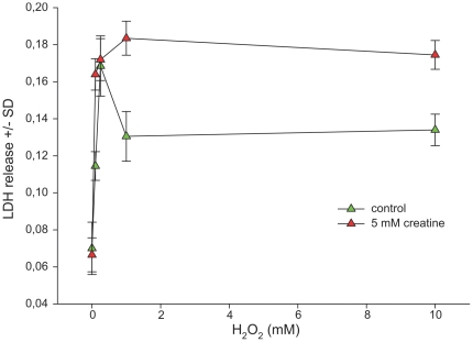
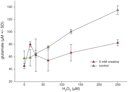
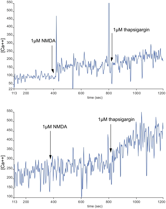
Creatine has been shown to be neuroprotective in aging, neurodegenerative conditions and brain injury. As a common molecular background, oxidative stress and disturbed cellular energy homeostasis are key aspects in these conditions. Moreover, in a recent report we could demonstrate a life-enhancing and health-promoting potential of creatine in rodents, mainly due to its neuroprotective action. In order to investigate the underlying pharmacology mediating these mainly neuroprotective properties of creatine, cultured primary embryonal hippocampal and cortical cells were challenged with glutamate or H(2)O(2). In good agreement with our in vivo data, creatine mediated a direct effect on the bioenergetic balance, leading to an enhanced cellular energy charge, thereby acting as a neuroprotectant. Moreover, creatine effectively antagonized the H(2)O(2)-induced ATP depletion and the excitotoxic response towards glutamate, while not directly acting as an antioxidant. Additionally, creatine mediated a direct inhibitory action on the NMDA receptor-mediated calcium response, which initiates the excitotoxic cascade. Even excessive concentrations of creatine had no neurotoxic effects, so that high-dose creatine supplementation as a health-promoting agent in specific pathological situations or as a primary prophylactic compound in risk populations seems feasible. In conclusion, we were able to demonstrate that the protective potential of creatine was primarily mediated by its impact on cellular energy metabolism and NMDA receptor function, along with reduced glutamate spillover, oxidative stress and subsequent excitotoxicity.
Conflict of interest statement
Figures
Similar articles
-
Creatine affords protection against glutamate-induced nitrosative and oxidative stress.Neurochem Int. 2016 May;95:4-14. doi: 10.1016/j.neuint.2016.01.002. Epub 2016 Jan 19. Neurochem Int. 2016. PMID: 26804444
-
Protective effect of the energy precursor creatine against toxicity of glutamate and beta-amyloid in rat hippocampal neurons.J Neurochem. 2000 May;74(5):1968-78. doi: 10.1046/j.1471-4159.2000.0741968.x. J Neurochem. 2000. PMID: 10800940
-
Clenbuterol protects mouse cerebral cortex and rat hippocampus from ischemic damage and attenuates glutamate neurotoxicity in cultured hippocampal neurons by induction of NGF.Brain Res. 1996 Apr 22;717(1-2):44-54. doi: 10.1016/0006-8993(95)01567-1. Brain Res. 1996. PMID: 8738252
-
Neuroprotective effects of creatine.Amino Acids. 2011 May;40(5):1305-13. doi: 10.1007/s00726-011-0851-0. Epub 2011 Mar 30. Amino Acids. 2011. PMID: 21448659 Review.
-
Creatine as a Neuroprotector: an Actor that Can Play Many Parts.Neurotox Res. 2019 Aug;36(2):411-423. doi: 10.1007/s12640-019-00053-7. Epub 2019 May 8. Neurotox Res. 2019. PMID: 31069754 Review.
Cited by
-
Diminution of Oxidative Damage to Human Erythrocytes and Lymphocytes by Creatine: Possible Role of Creatine in Blood.PLoS One. 2015 Nov 10;10(11):e0141975. doi: 10.1371/journal.pone.0141975. eCollection 2015. PLoS One. 2015. PMID: 26555819 Free PMC article.
-
In vivo neurometabolic profiling in patients with spinocerebellar ataxia types 1, 2, 3, and 7.Mov Disord. 2015 Apr 15;30(5):662-70. doi: 10.1002/mds.26181. Epub 2015 Mar 15. Mov Disord. 2015. PMID: 25773989 Free PMC article.
-
A guide to the metabolic pathways and function of metabolites observed in human brain 1H magnetic resonance spectra.Neurochem Res. 2014 Jan;39(1):1-36. doi: 10.1007/s11064-013-1199-5. Epub 2013 Nov 21. Neurochem Res. 2014. PMID: 24258018 Review.
-
Creatine supplementation and muscle-brain axis: a new possible mechanism?Front Nutr. 2025 Jul 23;12:1579204. doi: 10.3389/fnut.2025.1579204. eCollection 2025. Front Nutr. 2025. PMID: 40771202 Free PMC article. Review.
-
Effects of Creatine Supplementation on Brain Function and Health.Nutrients. 2022 Feb 22;14(5):921. doi: 10.3390/nu14050921. Nutrients. 2022. PMID: 35267907 Free PMC article. Review.
References
-
- Adcock KH, Nedelcu J, Loenneker T, Martin E, Wallimann T, et al. Neuroprotection of creatine supplementation in neonatal rats with transient cerebral hypoxia-ischemia. Dev Neurosci. 2002;24:382–388. - PubMed
-
- Beal MF. Mitochondria take center stage in aging and neurodegeneration. Ann Neurol. 2005;58:495–505. - PubMed
-
- Harman D. The biologic clock: the mitochondria? J Am Geriatr Soc. 1972;20:145–147. - PubMed
-
- Bender A, Krishnan KJ, Morris CM, Taylor GA, Reeve AK, et al. High levels of mitochondrial DNA deletions in substantia nigra neurons in aging and Parkinson disease. Nat Genet. 2006;38:515–517. - PubMed
MeSH terms
Substances
LinkOut - more resources
Full Text Sources
Other Literature Sources
Medical
Research Materials
