

Neonatal neurobehavior and diffusion MRI changes in brain reorganization due to intrauterine growth restriction in a rabbit model
- PMID: 22347486
- PMCID: PMC3275591
- DOI: 10.1371/journal.pone.0031497
Neonatal neurobehavior and diffusion MRI changes in brain reorganization due to intrauterine growth restriction in a rabbit model
Abstract
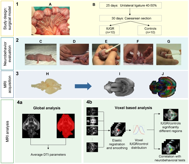
Background: Intrauterine growth restriction (IUGR) affects 5-10% of all newborns and is associated with a high risk of abnormal neurodevelopment. The timing and patterns of brain reorganization underlying IUGR are poorly documented. We developed a rabbit model of IUGR allowing neonatal neurobehavioral assessment and high resolution brain diffusion magnetic resonance imaging (MRI). The aim of the study was to describe the pattern and functional correlates of fetal brain reorganization induced by IUGR.
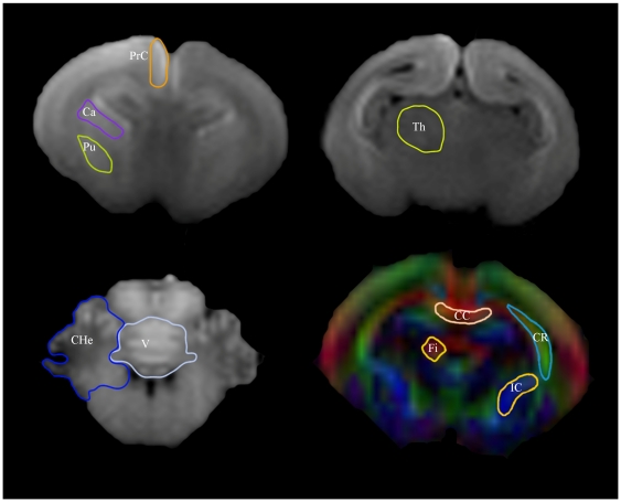
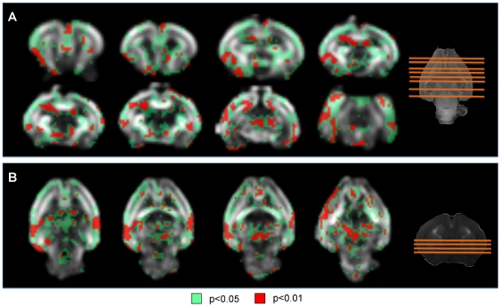
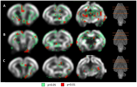
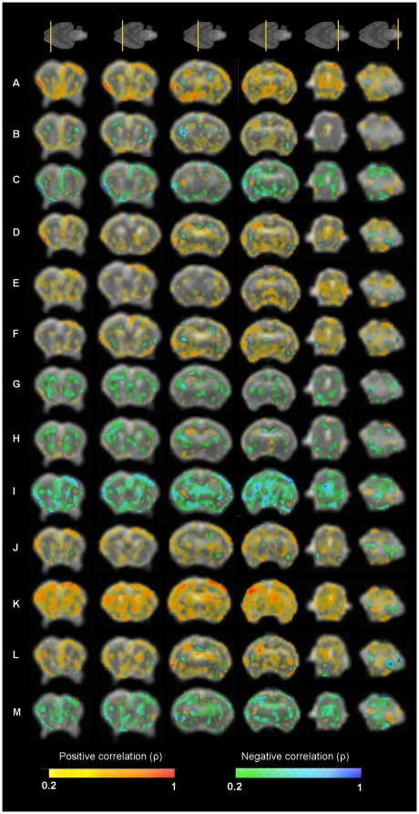
Methodology/principal findings: IUGR was induced in 10 New Zealand fetal rabbits by ligation of 40-50% of uteroplacental vessels in one horn at 25 days of gestation. Ten contralateral horn fetuses were used as controls. Cesarean section was performed at 30 days (term 31 days). At postnatal day +1, neonates were assessed by validated neurobehavioral tests including evaluation of tone, spontaneous locomotion, reflex motor activity, motor responses to olfactory stimuli, and coordination of suck and swallow. Subsequently, brains were collected and fixed and MRI was performed using a high resolution acquisition scheme. Global and regional (manual delineation and voxel based analysis) diffusion tensor imaging parameters were analyzed. IUGR was associated with significantly poorer neurobehavioral performance in most domains. Voxel based analysis revealed fractional anisotropy (FA) differences in multiple brain regions of gray and white matter, including frontal, insular, occipital and temporal cortex, hippocampus, putamen, thalamus, claustrum, medial septal nucleus, anterior commissure, internal capsule, fimbria of hippocampus, medial lemniscus and olfactory tract. Regional FA changes were correlated with poorer outcome in neurobehavioral tests.
Conclusions: IUGR is associated with a complex pattern of brain reorganization already at birth, which may open opportunities for early intervention. Diffusion MRI can offer suitable imaging biomarkers to characterize and monitor brain reorganization due to fetal diseases.
Conflict of interest statement
Figures
References
-
- Walker DM, Marlow N. Neurocognitive outcome following fetal growth restriction. Archives of Disease in Childhood- Fetal &Neonatal Edition. 2008;93:F322–325. - PubMed
-
- Baschat AA. Pathophysiology of fetal growth restriction: implications for diagnosis and surveillance. Obstet Gynecol Surv. 2004;59:617–627. - PubMed
-
- Rees S, Harding R, Walker D. An adverse intrauterine environment: implications for injury and altered development of the brain. Int J Dev Neurosci. 2008;26:3–11. - PubMed
-
- Bassan H, Stolar O, Geva R, Eshel R, Fattal-Valevski A, et al. Intrauterine growth-restricted neonates born at term or preterm: how different? Pediatric Neurology. 2011;44:122–130. - PubMed
-
- Figueras F, Oros D, Cruz-Martinez R, Padilla N, Hernandez-Andrade E, et al. Neurobehavior in term, small-for-gestational age infants with normal placental function. Pediatrics. 2009;124:e934–941. - PubMed
