

doi: 10.1155/2012/806989.
Epub 2012 Jan 24.
Anterior segment tomography with the cirrus optical coherence tomography
Affiliations
- PMID: 22347622
- PMCID: PMC3278920
- DOI: 10.1155/2012/806989
Item in Clipboard
Anterior segment tomography with the cirrus optical coherence tomography
J Ophthalmol.
2012.
Abstract
Optical coherence tomography (OCT) is an optical acquisition method to examine biological tissues. In recent years, OCT has become an important imaging technology used in diagnosing and following macular pathologies. Further development enabled application of optical coherence tomography in evaluation of the integrity of the nerve fiber layer, optic nerve cupping, anterior chamber angle, or corneal topography. In this manuscript we overview the use of OCT in the clinical practice to enable corneal, iris, ciliary body, and angle evaluation and diagnostics.
Figures
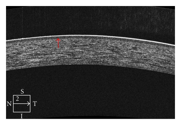
High-definition OCT of the cornea enables localization of the interface between the corneal stroma and epithelial/Bowman's layer.
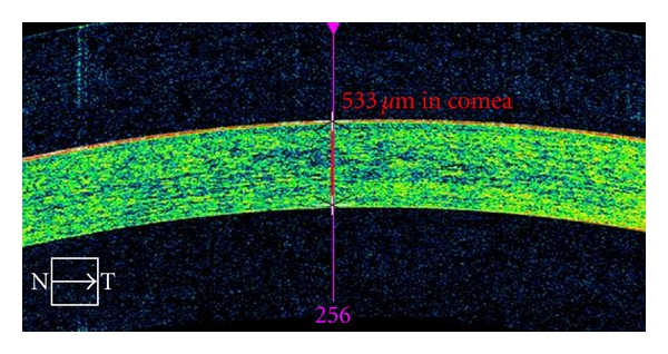
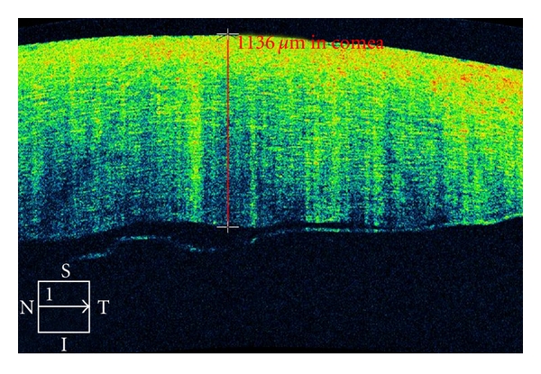
Anterior Segment Cube 512 × 128 imaging of the cornea enables pachymetry measurements.
Cirrus OCT of a 79-year-old patient who underwent cataract surgery. She developed postoperative corneal edema detected and monitored by the Cirrus OCT, characterized by diffuse hyperreflectivity interspacing lacunae of hyporeflectivity on corneal OCT scan. In addition, interruption of the endothelial layer can be documented.
Corneal imaging with the Cirrus OCT of an 86-year-old female patient who underwent lens phacoemulsification for therapy of dense nigra cataract. She developed focal microbullous keratopathy. OCT shows both the remarkable corneal edema, as well as interruption of endothelium layer. The patient was treated with topical steroids and experience improvement 4 weeks later.
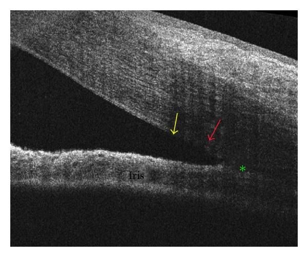
The Cirrus HD-OCT may enable identification of Schwalbe's line (yellow arrow), scleral spur (green asterisk), and trabecular meshwork (red arrow).
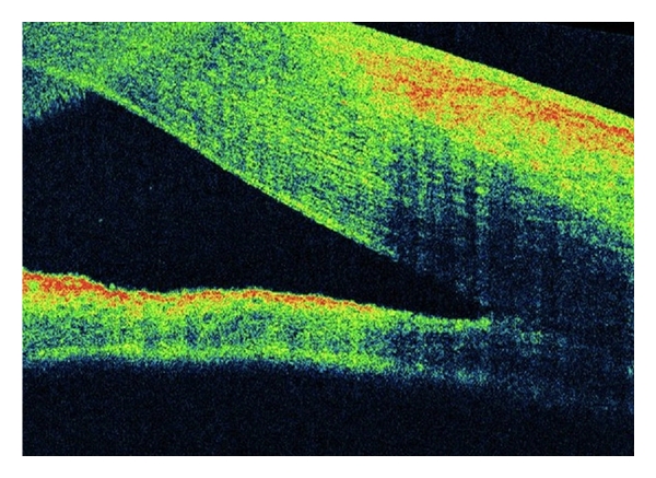
A 45-year-old female underwent vitrectomy plus silicone oil for retinal detachment repair. She presented with postoperative corneal edema that impaired angle examination. Cirrus OCT disclosed open anterior chamber angle.
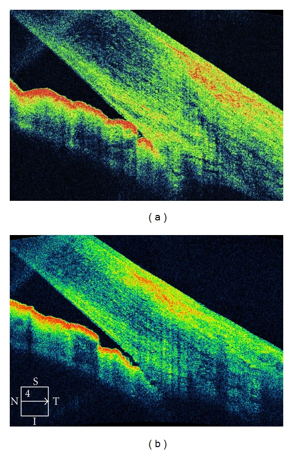
A 49-year-old female presents with a 1-week history of pain and visual loss OS. Ophthalmic examination was unremarkable, except for angle alterations and intraocular pressure of 57 mmHg OS. There were OS areas alternating very narrow and close anterior chamber angle; OD the angle was narrow. (a) OCT examination confirmed the anterior chamber angle morphology (b) The patient underwent topical and systemic antihypertensive ocular therapy. Two days later, the intraocular pressure was 16 mmHg OS, the angle was open, confirmed by OCT exam.
References
-
- Doors M, Berendschot TTJM, de Brabander J, Webers CAB, Nuijts RMMA. Value of optical coherence tomography for anterior segment surgery. Journal of Cataract and Refractive Surgery. 2010;36(7):1213–1229. - PubMed
-
- Simpson T, Fonn D. Optical coherence tomography of the anterior segment. Ocular Surface. 2008;6(3):117–127. - PubMed
-
- Izatt JA, Hee MR, Swanson EA, et al. Micrometer-scale resolution imaging of the anterior eye in vivo with optical coherence tomography. Archives of Ophthalmology. 1994;112(12):1584–1589. - PubMed
LinkOut - more resources
Full Text Sources
