. 2011:1:65.
doi: 10.4103/2156-7514.92187. Epub 2011 Dec 31.

Gardner's Syndrome

Affiliations

Gardner's Syndrome

Sapna Panjwani et al. J Clin Imaging Sci. 2011.

Abstract

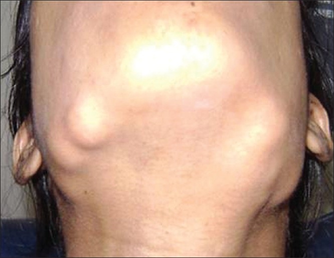
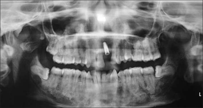
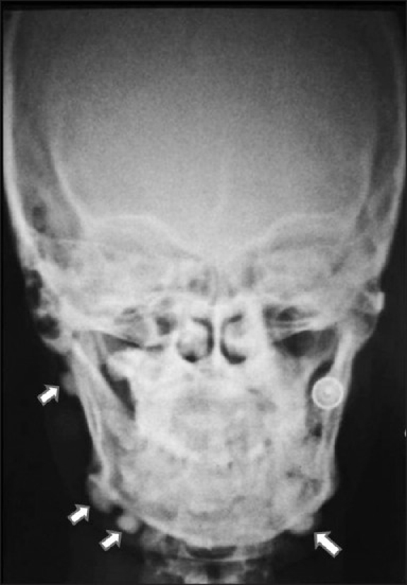
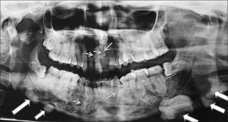
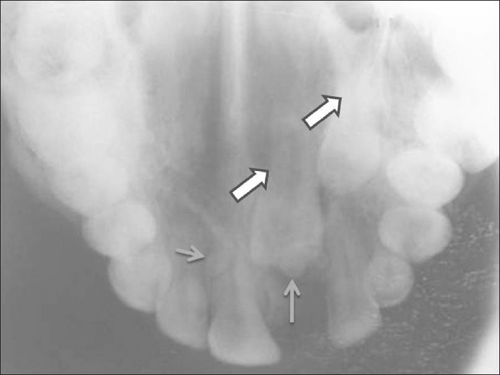
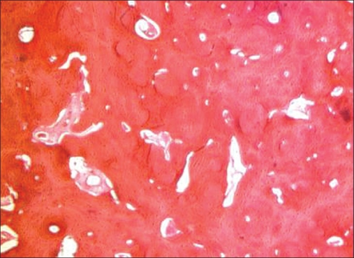
Gardner's syndrome is an autosomal dominant disease and is a subtype of familial adenomatous polyposis. It is characterized by adenomatous intestinal polyps, multiple osteomas in the skull, maxillae, mandible, and multiple cutaneous and subcutaneous masses (epidermoids and desmoid). Intestinal polyps, if not treated, have 100% chance of becoming malignant. We report a case of a 25-year-old female patient with Gardner's syndrome, with clinical manifestations including impacted supernumerary teeth, odontomes, sebaceous cyst on the scalp, and osteomas. It is important for the general dental practitioners to be aware of the clinical and radiological characteristics of Gardner's syndrome.

Keywords: Familial intestinal polyposis; gardner's syndrome; impacted teeth; osteoma.


Conflict of interest statement

Conflict of Interest: None declared.

Figures

Figure 1
Figure 1
Extraoral osteomas. A 25-year-old female patient with diffuse bilateral swellings on the lower third of face along the mandibular border.
Figure 2
Figure 2
Postoperative radiograph after removal of osteomas, impacted teeth, and implant placement, with respect to tooth 21.
Figure 3
Figure 3
Posteroanterior radiograph shows extra-oral osteomas along the mandible (white arrows).
Figure 4
Figure 4
OPG demonstrates impacted teeth 21 and 23, homogenous radiopaque masses over the right and left inferior aspect of the mandible suggestive of osteomas (large arrows). Also multiple small, homogeneous radiopaque masses surrounded by a radiolucent halo could be seen throughout the maxilla and mandible suggestive of complex odontomes (small arrows).Diffuse sclerotic masses are present throughout the body of mandible giving it a mottled appearance
Figure 5
Figure 5
Maxillary occlusal radiograph shows impacted teeth 21, 23 (large arrows) and odontomes (small arrows) in the near vicinity.
Figure 6
Figure 6
Photomicrograph shows the presence of compact lamellar bone with haversian canal, lacunae, histiocytes and reversal and resting lines suggestive of osteoma. (Hematoxylin-Eosin stain 10×).

Similar articles

Cited by

References

    1. Gardner EJ. “A genetic and clinical study of intestinal polyposis, a predisposing factor for carcinoma of the colon and rectum”. Am J Hum Genet. 1951;3:167–76. - PMC - PubMed
    1. Gu GL, Wang SL, Wei XM, Bai L. Diagnosis and treatment of Gardner's syndrome with gastric polyposis: A case report and review of literature. World J Gastroenterol. 2008;14:2121–3. - PMC - PubMed
    1. Lyons LA, Lewis RA, Strong LC, Zuckerbrod S, Ferrell RE. A Genetic study of Gardner syndrome and Congenital Hypertrophy of the Retinal Pigment Epithelium. Am J Hum Genet. 1988;42:290–6. - PMC - PubMed
    1. Davies AS. Gardner's syndrome: A case report. Br J Oral Surg. 1970;8:51–7. - PubMed
    1. Gorlin RJ, Pindborg JJ, Cohen MM. Syndromes of the Head and Neck. 2nd ed. New York: McGraw-Hill; 1976. pp. 324–8.