. 2012 Feb;8(2):e1002515.
doi: 10.1371/journal.pgen.1002515. Epub 2012 Feb 9.

Neurophysiological defects and neuronal gene deregulation in Drosophila mir-124 mutants

Affiliations

Neurophysiological defects and neuronal gene deregulation in Drosophila mir-124 mutants

Kailiang Sun et al. PLoS Genet. 2012 Feb.

Abstract

miR-124 is conserved in sequence and neuronal expression across the animal kingdom and is predicted to have hundreds of mRNA targets. Diverse defects in neural development and function were reported from miR-124 antisense studies in vertebrates, but a nematode knockout of mir-124 surprisingly lacked detectable phenotypes. To provide genetic insight from Drosophila, we deleted its single mir-124 locus and found that it is dispensable for gross aspects of neural specification and differentiation. On the other hand, we detected a variety of mutant phenotypes that were rescuable by a mir-124 genomic transgene, including short lifespan, increased dendrite variation, impaired larval locomotion, and aberrant synaptic release at the NMJ. These phenotypes reflect extensive requirements of miR-124 even under optimal culture conditions. Comparison of the transcriptomes of cells from wild-type and mir-124 mutant animals, purified on the basis of mir-124 promoter activity, revealed broad upregulation of direct miR-124 targets. However, in contrast to the proposed mutual exclusion model for miR-124 function, its functional targets were relatively highly expressed in miR-124-expressing cells and were not enriched in genes annotated with epidermal expression. A notable aspect of the direct miR-124 network was coordinate targeting of five positive components in the retrograde BMP signaling pathway, whose activation in neurons increases synaptic release at the NMJ, similar to mir-124 mutants. Derepression of the direct miR-124 target network also had many secondary effects, including over-activity of other post-transcriptional repressors and a net incomplete transition from a neuroblast to a neuronal gene expression signature. Altogether, these studies demonstrate complex consequences of miR-124 loss on neural gene expression and neurophysiology.


Conflict of interest statement

The authors have declared that no competing interests exist.

Figures
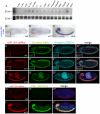
Figure 1
Figure 1. Temporal and spatial expression of Drosophila miR-124.
(A) Northern analysis using staged preparations of total RNA. (B–D) Nascent transcription of pri-mir-124 detected with a 1 kb probe. (B and C) are ventral views and (D) is a lateral view. Inset of panel (B) highlights the detection of nuclear dots that reflect the chromosomal locations of mir-124 transcription. (E–I) Expression of a miR-124:DsRed transgene colabeled with various neural markers, Deadpan (neuroblast marker) and Prospero (ganglion mother cell marker) and Elav (differentiated neuron marker); embryos in H–I are counterstained with DAPI. In all panels, miR-124:DsRed is at left, the neural markers in the middle, and merged images at right; the signal in the center of panels G is gut autofluorescence. Activity of mir-124 initiates in neuroblasts and is maintained in GMCs and CNS neurons. High magnification insets of panels E–F show gradual expression of miR-124:DsRed in all Deadpan+ and Prospero+ positive cells in CNS.
Figure 2
Figure 2. General characterization of mir-124 knockout and rescue strains.
(A) The pre-mir-124 hairpin was replaced with mini-white+ using ends-out homologous recombination. A 19 kb mir-124 rescue transgene lacks known protein-coding genes; it overlaps mir-287 but this locus has not been validated as a miRNA from deep sequencing . (B) Northern validation that adult mir-124 knockouts are null for mature miR-124; a normal level of miR-124 is restored by the rescue transgene. (C) The substantially shortened lifespan of mir-124 transheterozygous males raised at 29°C was fully rescued by the mir-124 transgene.
Figure 3
Figure 3. Absence of major defects in specification of the nervous system of mir-124 mutants.
(A–F) Stage ∼13 embryos, ventral aspect. A and B are triple labelings of Deadpan (NB), Prospero (GMC) and Elav (neuron); no substantial differences were observed. (C) Graph indicates the number of Dpn+ cells per hemisegment comparing mir124[6/6] embryos to mutants carrying the genomic rescue. Error bar represents standard deviation from the average of five embryos; 20 and 30 hemisegments were quantified for the thoracic and abdominal segments, respectively. Since subtler differences might not be seen with pan-neuronal labeling, we analyzed Eve (D, E), which is active in a subset of CNS neurons and sibling cells; the mutant was similar to wildtype. (F, G) Expression of the glial marker Repo was not markedly different in mir-124 mutants. (H–J) Larval brains. (H–I) Specification of neuroblasts and neurons is relatively similar in wildtype and mir-124 mutant. (J) MARCM analysis in mir-124 mutant brain to mark the lineages produced by single neuroblasts. GFP+ mutant clones maintain a single neuroblast (marked by large Dpn+ cells, arrows in J″) and can generate multiple neurons.
Figure 4
Figure 4. Requirement of mir-124 for larval locomotion and synaptic transmission.
(A–C) Locomotion defects in mir-124 mutants. Each track depicts the movement of an individual 3rd instar larva tracked for one minute; 15 tracks were superimposed to reveal population behavior. (D) Quantitative analysis showed that both mir-124 mutant genotypes exhibited substantially reduced locomotion, which was rescued by mir-124 genomic transgene. (E, F) HRP staining (green) of the neuromuscular junction on muscle 4 (counterstained in phalloidin) in wildtype and mir-124 mutant. (G) Quantitative analysis (n = 12) showed no significant difference in bouton numbers or NMJ area, as normalized to muscle surface area (MSA). (H–K) Loss of miR-124 leads to an enhancement of presynaptic neurotransmitter release. (H–J) Representative traces of evoked (excitatory junction currents, EJC) and spontaneous (miniature EJC, mEJC) membrane currents recorded from muscle 6 in the third abdominal segment in wandering third-instar larvae of w[1118]; mir-124[12/12] and mir-124[12/12] rescued by genomic insert. EJC contain 10 consecutive superimposed traces and mEJC are three traces of continuous recordings. (K) Quantification of mEJC, EJC, and quantal content (QC) for the indicated genotypes. Deletion of mir-124 resulted in no differences in spontaneous activity, but caused significant increases in evoked currents and quantal content; these phenotypes were rescuable. n = 22 NMJs for each genotype. Error bars represent SEM, statistical tests by two-tailed t-test show *p<0.05, **p<0.01, ***p<0.001.
Figure 5
Figure 5. mir-124 suppresses variation in dendrite numbers on sensory neurons.
(A–C) We labeled ddaD and ddaE neurons with CD8-GFP under the control of Gal4221. Representative images are shown from mir-124 loss- and gain-of-function backgrounds. The overall patterns of dendrite branching in wildtype (A) and mutant (B) were similar. (C) Misexpression of mir-124 in the mir-124 mutant strongly decreased the complexity of dendritic branching. (D) Quantitative analysis. Each of the circles represents dendrite quantification of an individual neuron. Although the average numbers of dendritic ends for ddaD and ddaE neurons in mir-124[6] or mir-124[7] mutants were not statistically different those in wildtype, the variation in their numbers was significantly increased (by F test). The effect was more pronounced in ddaD than ddaE neurons, but both were rescued by the mir-124 genomic transgene. Analysis of mir-124 overexpression in the mir-124 mutant background showed a strong decrease in branching complexity; still, the variation in dendritic end numbers was rescued.
Figure 6
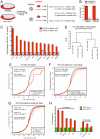
Figure 6. Gene expression in miR-124+ cells from wild-type and mir-124 mutants.
(A) Scheme for isolation and analysis of cells. (B) qPCR validation that the sorted DsRed+ cells specifically express mir-124. (C) qPCR analysis of predicted miR-124 targets showed upregulation in miR-124:DsRed+ cells, but not miR-124:DsRed- cells. (D) Unsupervised hierarchical clustering of microarray data from miR-124:DsRed+ cells purified from wildtype and mir-124 mutant embryos collected 10–16 hrs after egg laying. (E–G) Cumulative distribution function (CDF) plots of various sets of predicted miR-124 targets in mutant vs. wildtype microarray data. Shifts to the right reflect overall upregulation of genes in the mir-124 mutant. (E) Global upregulation of all predicted miR-124 targets. (F) Transcripts with well-conserved miR-124 sites were upregulated more strongly than those with poorly-conserved target sites, although both sets were significantly upregulated. (G) Transcripts with 2–8 seed matches were upregulated more strongly than transcripts with 2–7 or non-canonical seed matches. (H) Sensor validation of direct repression of miR-124 targets by ectopic miR-124. Transcripts with 2–8 (7mer) targets generally repressed more strongly than those with 2–7 (6mer sites).
Figure 7
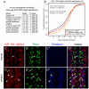
Figure 7. Lack of evidence for mutual exclusion amongst the functional miR-124 target network.
(A) Consistent with earlier reports , transcripts bearing miR-124 target sites predicted by mirSVR are enriched for genes annotated with non-neural expression. The top enriched tissue annotations are shown in rank order. (B) The absolute levels of transcripts bearing miR-124 target sites in miR-124:DsRed+ cells are well above average gene expression. Moreover, transcripts bearing well-conserved sites are overall more highly expressed than those with poorly-conserved sites. (C, D) Triple label of stage 14 embryos for miR-124:DsRed, Repo (a glial marker) and Deadpan (a neuroblast marker). (C) In more dorsal planes lacking neuroblasts, miR-124:DsRed is excluded from most Repo+ cells, although rare cells show colocalization (arrow). (D) In more ventral planes containing NBs, miR-124:DsRed colocalizes with Deadpan+ cells (arrowheads) as well as Repo+ (arrows) cells inferred to be glioblasts.
Figure 8
Figure 8. Functional interpretation of direct and indirect consequences of miR-124 loss.
(A) Core components of the retrograde BMP signaling pathway at the NMJ. Release of the glass bottom boat (Gbb) ligand from the muscle activates BMP receptors (Sax, Tkv and Wit) in the neuron. Activated BMP receptors induce phosphorylation of Mad, which partners with Medea to activate target genes, such as trio. (B) miR-124 binding sites; HC = highly conserved and PC = poorly conserved (see also Figure S8). (C) Sensor assays in S2 cells confirm that the 3′ UTRs of all five BMP pathway components are responsive to miR-124. (D–F) Ectopic activation of Tkv receptor can phenocopy mir-124 mutant electrophysiology. Representative traces of evoked (excitatory junction currents, EJC) and spontaneous (miniature EJC, mEJC) membrane currents recorded from muscle 6 in the third abdominal segment in w[1118]; BG380-Gal4/+ (D) and w[1118]; BG380-Gal4/+; UAS-TkvA/+ (E) wandering third-instar larvae. EJC contain 10 consecutive superimposed traces and mEJC are three traces of continuous recordings. (F) Quantification of mEJC, EJC, and quantal content (QC) for the indicated genotypes. Activated Tkv did not affect spontaneous activity, but caused significant increases in evoked currents and quantal content. n = 12 NMJs for each genotype. Error bars represent SEM, statistical tests by two-tailed t-test show *p<0.05. (G) miREDUCE analysis shows that variations of the miR-124 seed are strongly enriched amongst transcripts that increase in mir-124 mutant cells (highlighted in red). Amongst motifs associated with decreased gene expression in mir-124 mutants, the top motif corresponds to the Pumilio site; others motifs include the seeds of K box family miRNAs, miR-10-5p, and an orphan motif (AUGCAAA) with several hundred conserved matches (defined by TargetScan). (H) Cumulative distribution function (CDF) plots of gene in the neuron and NB clusters. The group of neural genes is shifted towards lower expression levels in the mir-124 mutant, while the NB cluster is shifted towards higher expression.

Similar articles

Cited by

References

    1. Lai EC. microRNAs are complementary to 3′ UTR sequence motifs that mediate negative post-transcriptional regulation. Nat Genet. 2002;30:363–364. - PubMed
    1. Doench JG, Sharp PA. Specificity of microRNA target selection in translational repression. Genes Dev. 2004;18:504–511. - PMC - PubMed
    1. Brennecke J, Stark A, Russell RB, Cohen SM. Principles of MicroRNA-Target Recognition. PLoS Biol. 2005;3:e85. doi: 10.1371/journal.pbio.0030085. - DOI - PMC - PubMed
    1. Lewis BP, Burge CB, Bartel DP. Conserved seed pairing, often flanked by adenosines, indicates that thousands of human genes are microRNA targets. Cell. 2005;120:15–20. - PubMed
    1. Friedman RC, Farh KK, Burge CB, Bartel DP. Most mammalian mRNAs are conserved targets of microRNAs. Genome Res. 2009;19:92–105. - PMC - PubMed

Publication types

LinkOut - more resources