. 2010 May;1(2):53-61.
doi: 10.1007/s13244-010-0017-8. Epub 2010 Apr 14.

Computed tomography of complicated Meckel's diverticulum in adults: a pictorial review

Computed tomography of complicated Meckel's diverticulum in adults: a pictorial review

Alexandra Platon et al. Insights Imaging. 2010 May.

Abstract

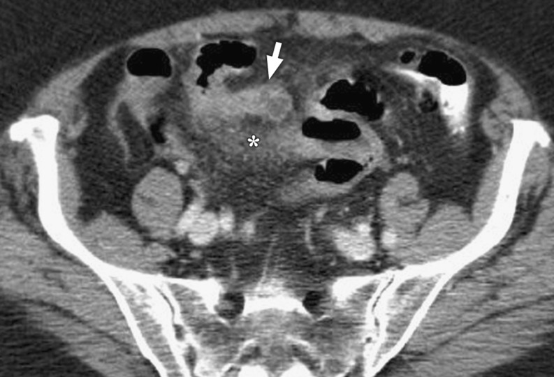
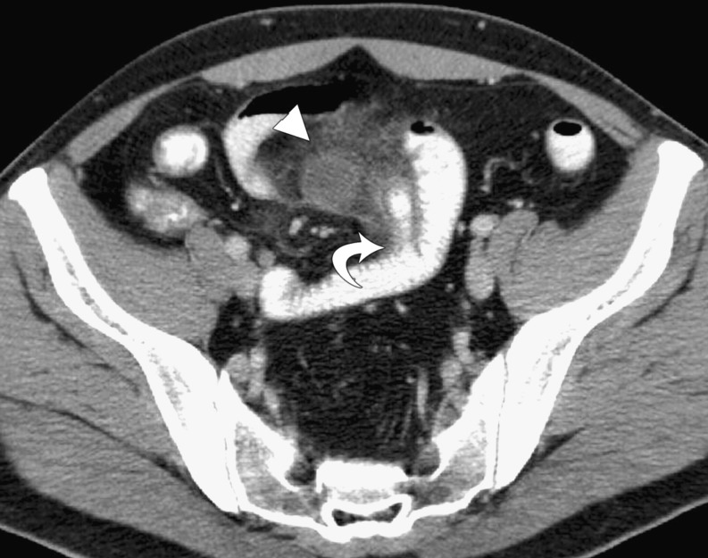
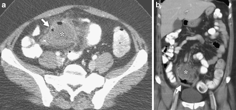
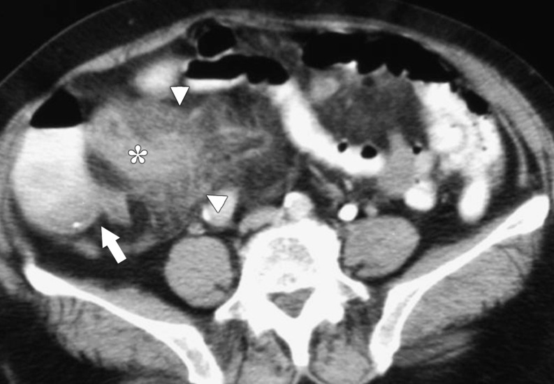
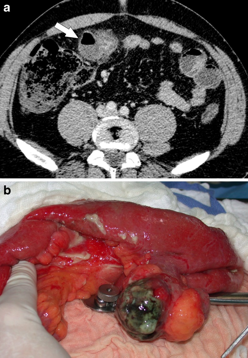
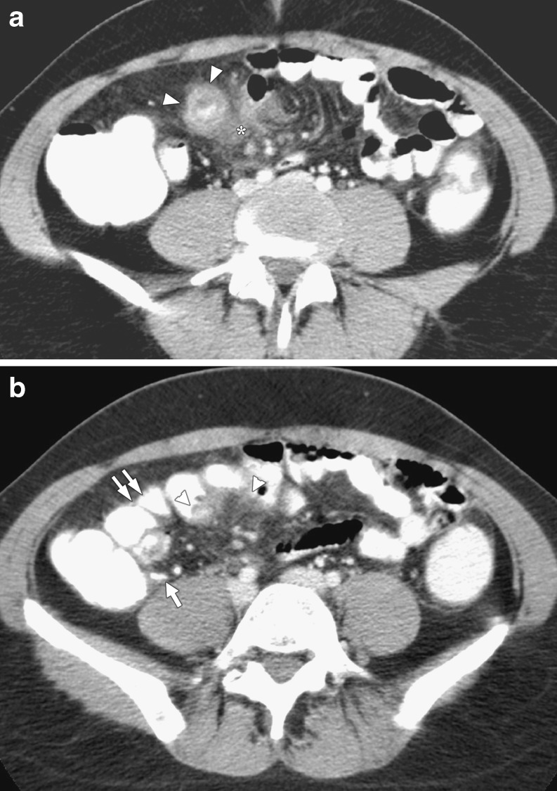
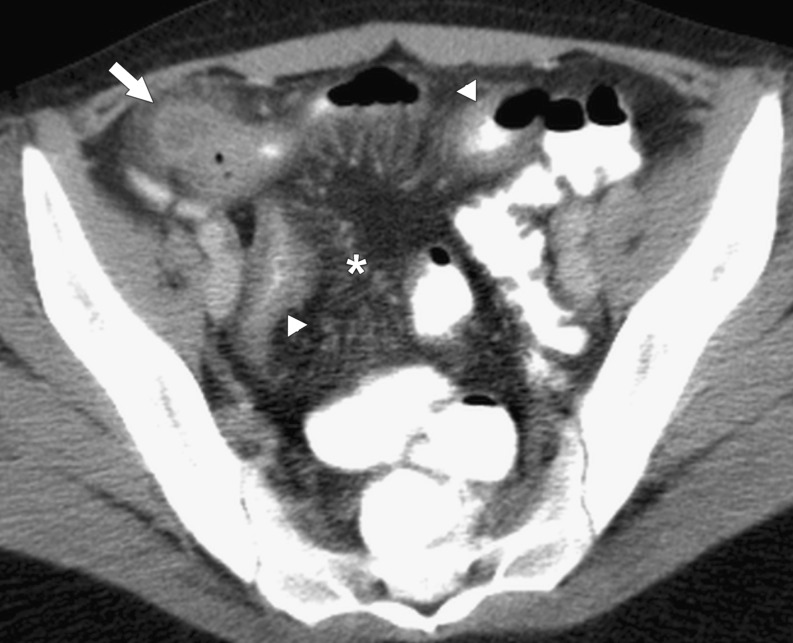
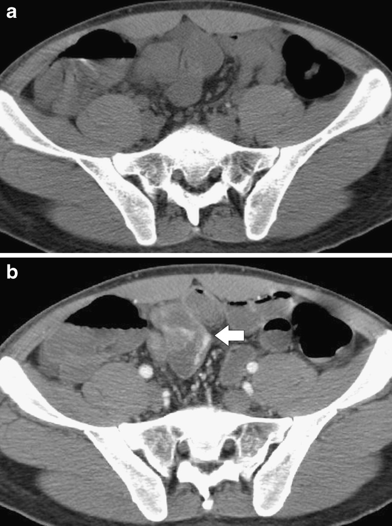
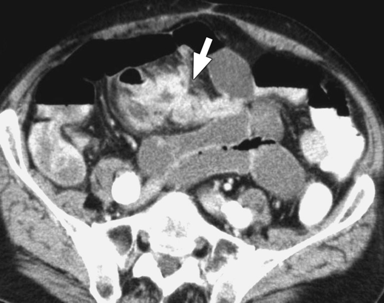
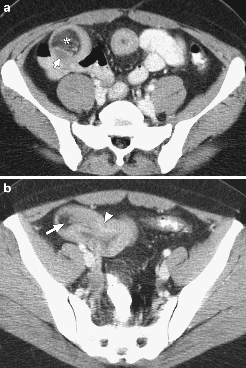
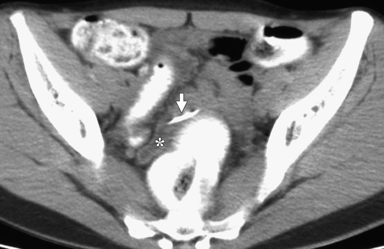
OBJECTIVE: To show various CT aspects of complicated Meckel's diverticulum in adult patients to facilitate the preoperative diagnosis of this rare pathology in emergency settings. METHODS: A computer search of medical records over a 15 year period identified 23 adult patients who underwent surgery for acute abdomen generated by a complicated Meckel's diverticulum. CT images available for review were analyzed, and some specific patterns leading to the diagnosis of complicated Meckel's diverticulum are presented in this review. RESULTS: Complications were related to inflammation (14 patients), bleeding (5 patients), intestinal obstruction (3 patients), and penetrating foreign body (1 patient). The presence of a Meckel's diverticulum was usually suggested at CT scan by an abnormal outpouching, blind-ending digestive structure connected to the terminal ileum by a neck of variable caliber. Depending on the type of complications, the diverticulum was surrounded by mesenteric inflammatory changes, or presented as a localized fluid or air-fluid collection contiguous with the terminal ileum. The diverticulum was also the source of active bleeding or acted as the lead point to intestinal obstruction or intussusception. CONCLUSION: CT findings of complicated Meckel's diverticulum are polymorphic and should be considered in the evaluation of adult patients with acute abdomen.


Figures

Fig. 1
Fig. 1
A 76-year-old patient with surgically proven Meckel’s diverticulitis. Intravenous contrast-enhanced CT shows the Meckel’s diverticulum as a fluid-filled, blind-ending digestive structure (arrow), arising from a distal ileal loop, along with peridiverticular mesenteric fat stranding (asterisk), suggestive of inflammation.
Fig. 2
Fig. 2
A 37-year-old man with inflamed Meckel’s diverticulum and abscess formation (surgery proven). Intravenous- and oral-enhanced CT scan shows a contrast-filled tubular diverticulum with a “mucosal triangular plateau” pattern at its ileal attachment (curved arrow); there is a fluid-filled collection at the diverticulum’s tip and associated fat stranding (arrowhead).
Fig. 3a, b
Fig. 3a, b
A 33-year-old woman with surgically proven perforated Meckel’s diverticulum. a Intravenous- and oral-enhanced CT shows the perforated Meckel’s diverticulum as a gas-containing collection in the right lower quadrant (asterisk), situated between the ileal loops and surrounded by fat stranding (arrow). b Coronal reconstructed CT demonstrates the perforated diverticulum (asterisk), contiguous with a thickened ileal loop (arrow).
Fig. 4
Fig. 4
A 71-year-old woman with surgically proven inflamed Meckel’s diverticulum with tip perforation. Intravenous- and oral-enhanced CT shows the diverticulum as a tubular, thickened-wall, blind-ending digestive structure (asterisk) surrounded by an important fat infiltration (arrowheads); the diverticulum is situated in the right lower quadrant. Caecum and terminal ileum are normal (arrow).
Fig. 5a, b
Fig. 5a, b
A 44-year-old man with surgically proven necrotic Meckel’s diverticulum (images courtesy of Dr. K. Bider and Dr. C. Beynon). a Intravenous-enhanced CT shows the inflamed diverticulum as an air-fluid collection (arrow) with thin, nonenhancing wall adjacent to an ileal loop. b Photograph taken during surgery before opening the specimen shows a 7 cm large, necrotic Meckel’s diverticulum.
Fig. 6a, b
Fig. 6a, b
A 32-year-old woman with surgically proven Meckel’s diverticulitis. a Intravenous- and oral-enhanced CT shows the inflamed diverticulum as a round, contrast-filled, thickened-wall collection (arrowheads) in continuation with an ileal loop; associated fat stranding (asterisk) is also demonstrated. b CT scan at a higher level than a shows the peridiverticular inflammatory changes (arrowheads) along with normal aspect of the appendix (arrow) and of the ileal loops proximal to the inflamed diverticulum (double arrows).
Fig. 7
Fig. 7
A 22-year-old woman with surgically proven perforated Meckel’s diverticulitis and Crohn’s disease. Intravenous- and oral-enhanced CT shows the collection due to the perforated Meckel’s diverticulum (arrow) in the right lower quadrant. Thickened-wall ileal loops (arrowheads) and prominently dilated mesenteric vessels (asterisk), proving active Crohn’s disease are also demonstrated.
Fig. 8a, b
Fig. 8a, b
A 22-year-old man with massive gastrointestinal bleeding due to heterotopic gastric mucosa in a Meckel’s diverticulum (surgery proven). a Nonenhanced CT scan at the same level as b shows no spontaneously dense material in the bowel lumen. b Intravenous-enhanced CT scan shows contrast extravasation seen as linear hyperattenuating pooling of contrast at the neck of the Meckel’s diverticulum (arrow), arising from a distal ileal loop.
Fig. 9
Fig. 9
A 63-year-old patient with small bowel obstruction due to inflammation of the Meckel’s diverticulum (surgery proven). Intravenous abdominal CT shows the diverticulum as an enhancing, blind-ending digestive structure, with thick wall and fluid content (arrow); this finding was demonstrated to be an inflamed Meckel’s diverticulum situated at the transition point with the dilated small bowel loops and surrounded by inflammatory changes in the mesenteric fat.
Fig. 10a, b
Fig. 10a, b
A 39-year-old woman with surgically proven ileal intussusception related to the Meckel’s diverticulum as the lead point. a Intravenous- and oral-enhanced CT scan shows the intussusception of the ileum (arrow) along with its mesentery (asterisk), creating the “target sign.” b Image at a lower level than a demonstrates the invaginated mesenteric fat (arrow), along with thickening of the intussuscepted ileum (arrowhead).
Fig. 11
Fig. 11
A 20-year-old woman with surgically proven needle retention in a giant Meckel’s diverticulum. Oral-enhanced CT shows the needle (arrow) located at the collar of a fluid-filled, blind-ended tubular structure (asterisk) that is the Meckel’s diverticulum.

Similar articles

Cited by

References

    1. Stone PA, Hofeldt MJ, Campbell JE, Vedula G, DeLuca JA, Flaherty SK. Meckel diverticulum: ten-year experience in adults. South Med J. 2004;97:1038–1041. doi: 10.1097/01.SMJ.0000125222.90696.03. - DOI - PubMed
    1. Levy AD, Hobbs CM. Meckel diverticulum: radiologic features with pathologic correlation. Radiographics. 2004;24:565–587. doi: 10.1148/rg.242035187. - DOI - PubMed
    1. Cullen JJ, Kelly KA, Moir CR, Hodge DO, Zinsmeister AR, Melton LJ., 3rd Surgical management of Meckel's diverticulum. An epidemiologic, population-based study. Ann Surg. 1994;220:564–568. doi: 10.1097/00000658-199410000-00014. - DOI - PMC - PubMed
    1. Park JJ, Wolff BG, Tollefson MK, Walsh EE, Larson DR. Meckel diverticulum: the Mayo Clinic experience with 1476 patients (1950–2002) Ann Surg. 2005;241:529–533. doi: 10.1097/01.sla.0000154270.14308.5f. - DOI - PMC - PubMed
    1. Rossi P, Gourtsoyiannis N, Bezzi M, Raptopoulos V, Massa R, Capanna G, Pedicini V, Coe M. Meckel's diverticulum: imaging diagnosis. AJR Am J Roentgenol. 1996;166:567–573. - PubMed

LinkOut - more resources