. 2010 Jul;1(3):155-166.
doi: 10.1007/s13244-010-0030-y. Epub 2010 Jul 27.

Flying through congested airspaces: imaging of chronic rhinosinusitis

Affiliations

Flying through congested airspaces: imaging of chronic rhinosinusitis

Davide Farina et al. Insights Imaging. 2010 Jul.

Abstract

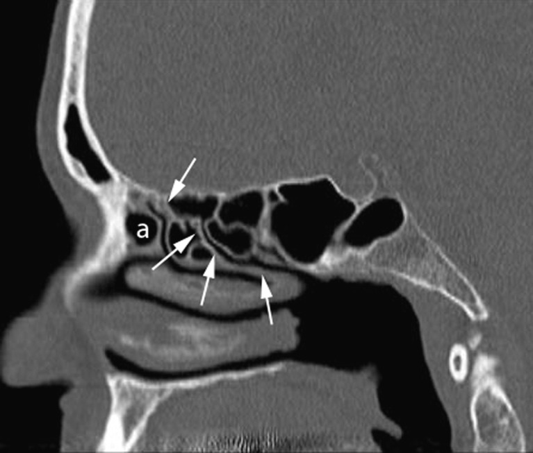
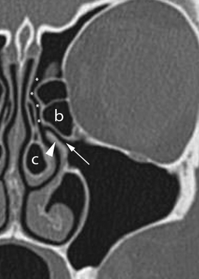
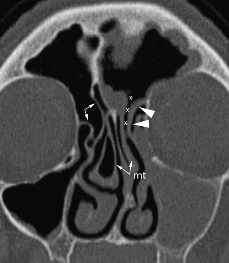
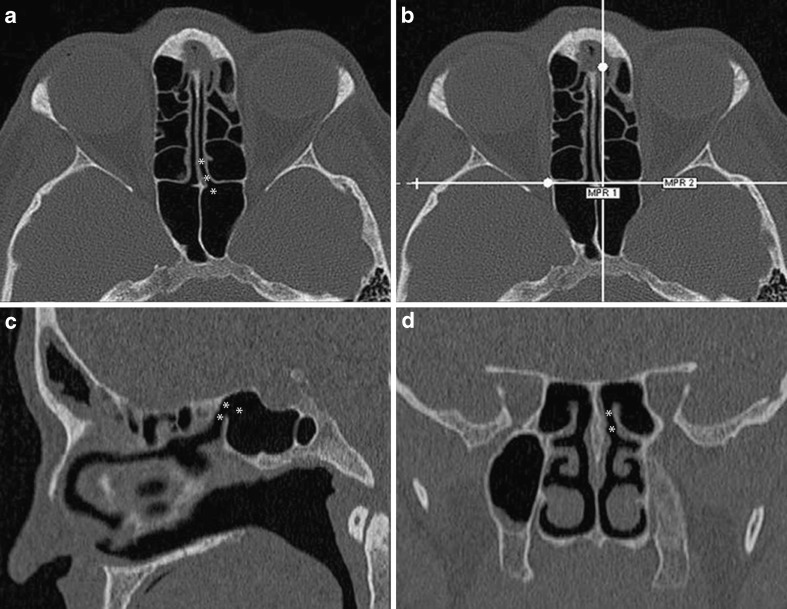
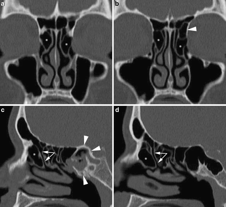
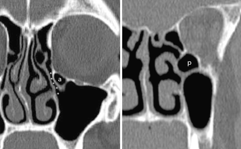
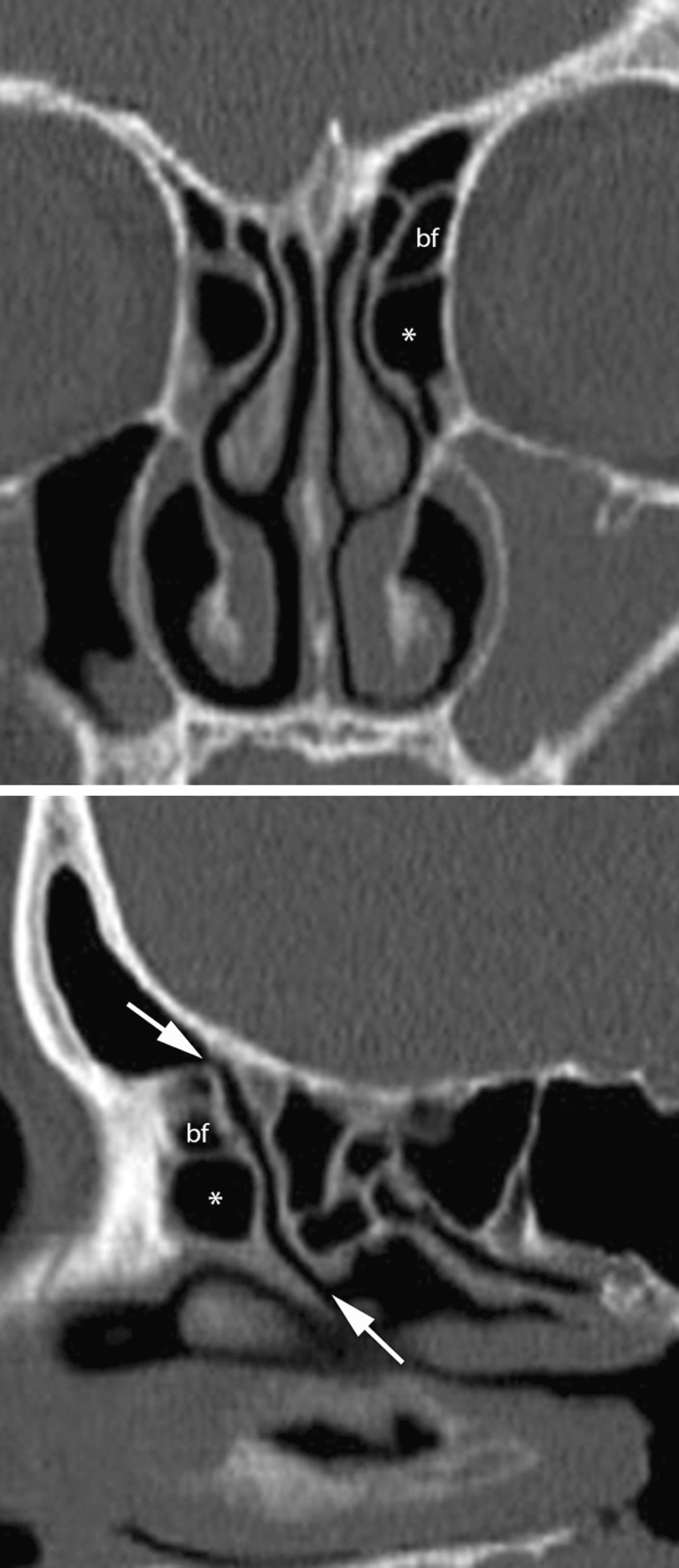
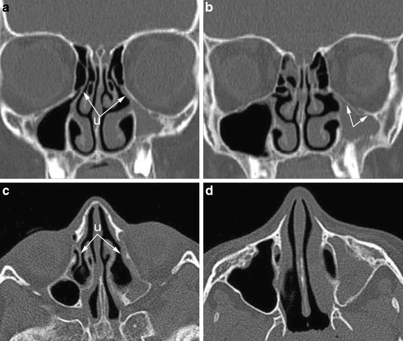
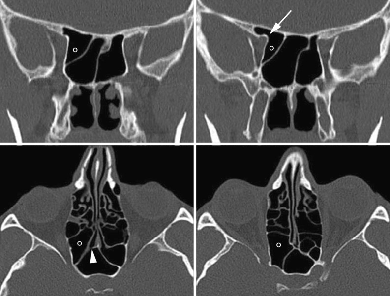
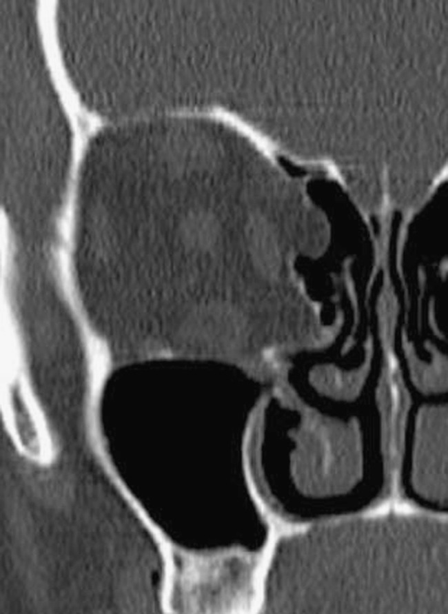
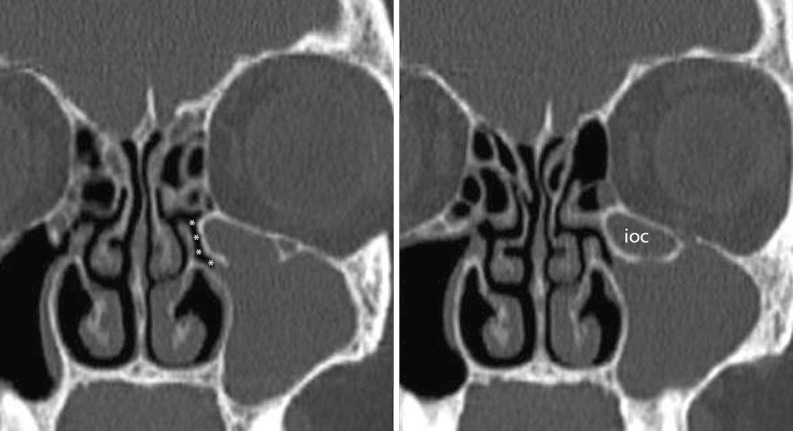
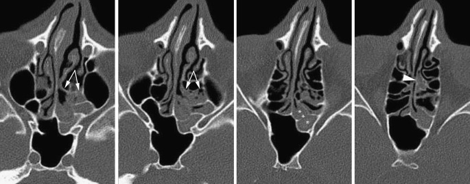
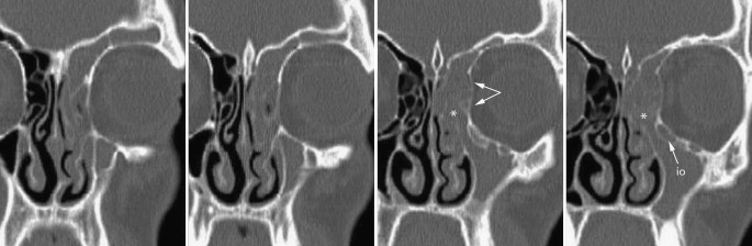
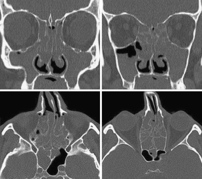
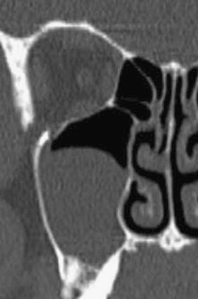
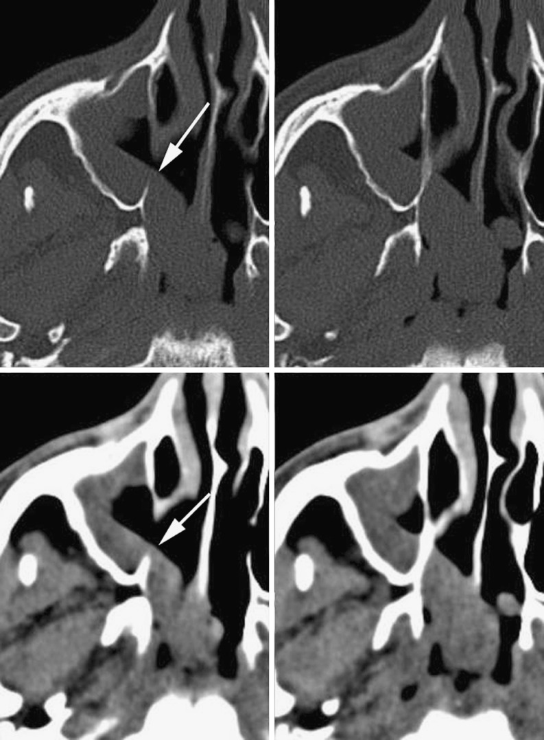
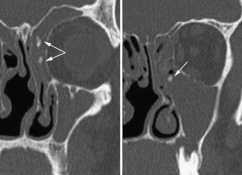
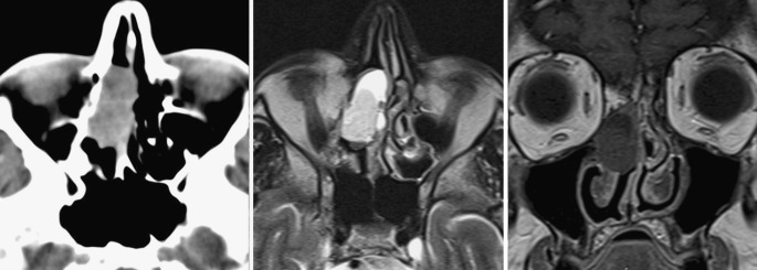
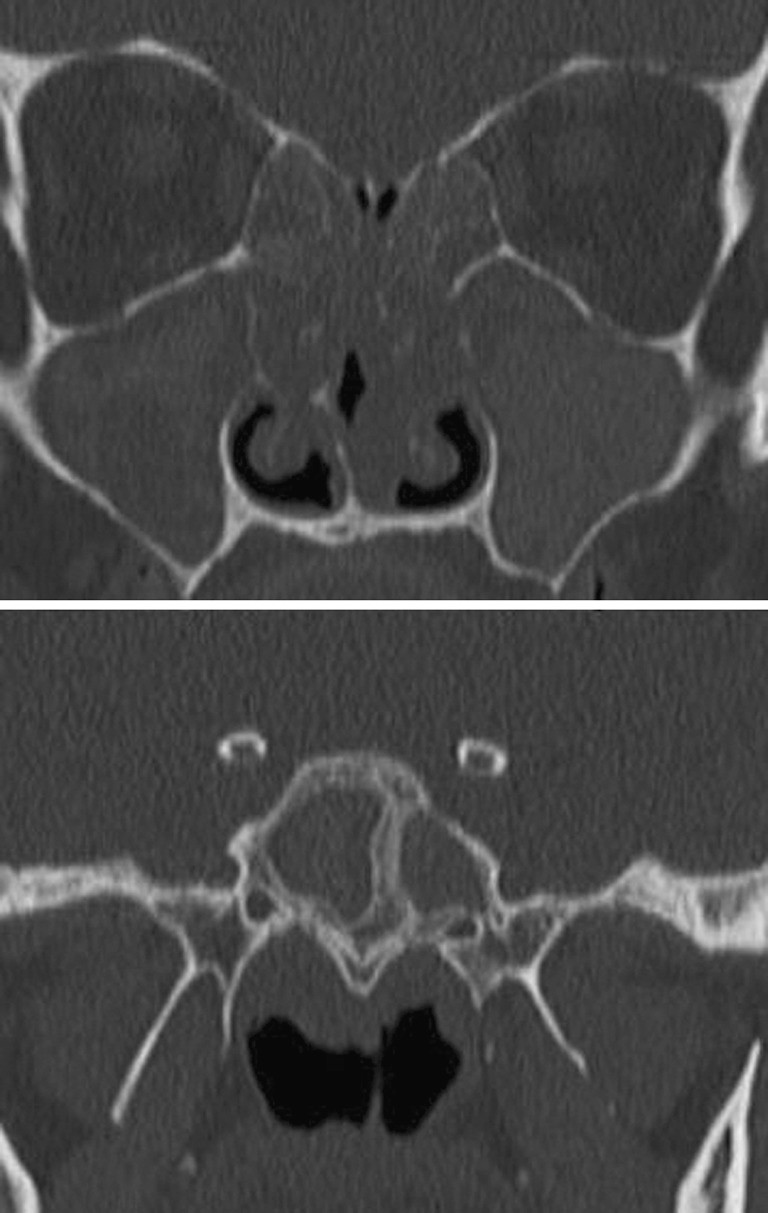
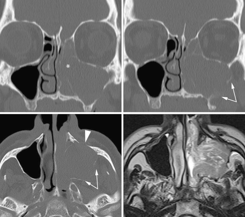
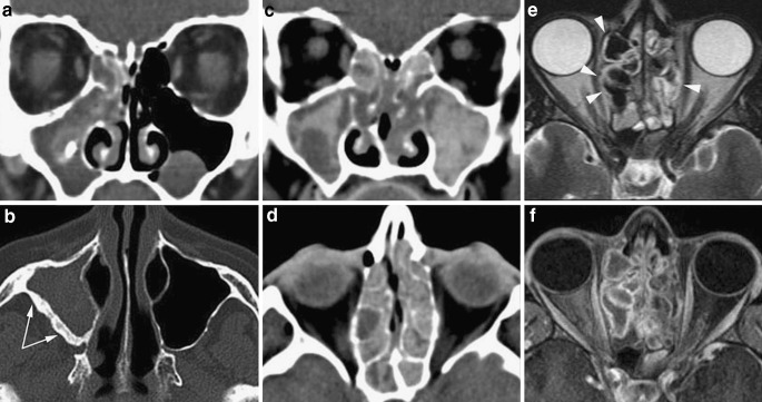
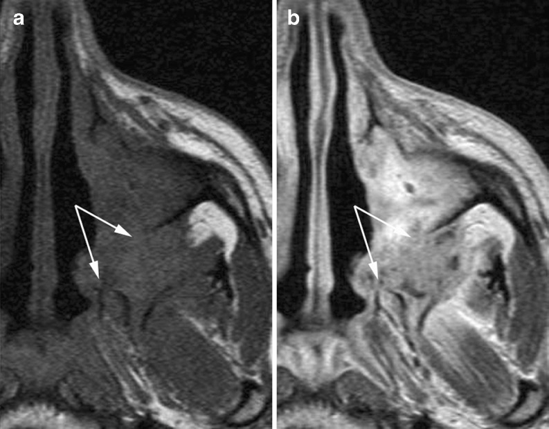
The complex regional anatomy of the nose and paranasal sinuses makes the interpretation of imaging studies of these structures intimidating to many radiologists. This paper aims to provide a key to interpretation by presenting a simplified approach to the functional anatomy of the paranasal sinuses and their most common (and most relevant) variants. This knowledge is basic for the full understanding of chronic rhinosinusitis and its computed tomography (CT) patterns. As fungal infections may be observed in the setting of chronic rhinosinusitis, these are also discussed. Chronic sinus inflammation produces bone changes, clearly depicted on CT images. Finally, clues to suspecting neoplastic lesions underlying inflammatory sinus conditions are provided.


Figures

Fig. 1
Fig. 1
Sagittal MPR reconstruction. Arrows outline the course of the basal lamella of the middle turbinate, which marks the border between anterior and posterior ethmoid air cells (a agger nasi cell)
Fig. 2
Fig. 2
Coronal MSCT. The ostiomeatal complex is the functional area in which the anterior ethmoid cells, and the maxillary and frontal sinus drain. It is composed of the middle turbinate (pneumatised in this case, c), the uncinate process (arrowhead), the ethmoid bulla (b), the ethmoid infundibulum (arrow), and the frontal recess (asterisks)
Fig. 3
Fig. 3
Coronal oblique MPR reconstruction showing the variable cranial attachment of the uncinate process. On the left side, the uncinate process attaches laterally to the medial orbital wall (arrowheads), thus the frontal recess (asterisks) courses close to the middle turbinate (mt). The ethmoid infundibulum is obstructed resulting in sinusitis with an infundibular pattern. On the right side, the uncinate process inserts on both the medial orbital wall and the skull base (arrows); the frontal recess (not visible) will drain into the middle meatus
Fig. 4
Fig. 4
The sphenoethmoid recess (asterisks) is generally well seen on the axial plane along the medial aspect of the anterior sphenoid sinus wall. Identification on the coronal and sagittal planes can be simplified navigating through the volume as shown in b, c and d
Fig. 5
Fig. 5
MSCT coronal (a, b) and sagittal reformations obtained on the right (c) and left (d) sides. Bilateral agger nasi cells (asterisks) are visible on the coronal images, but sagittal reformations better display their relationships with the frontal recesses (arrows). A type I bulla frontalis (arrowhead in b) can be seen on top of the left agger nasi cell. Inflammatory material with focal calcifications is retained within the right sphenoid sinus whose walls are densely sclerotic (arrowheads in c): chronic inflammation, possibly a fungus ball (see below)
Fig. 6
Fig. 6
Anterior (a) and posterior (p) infraorbital cells seen in two different patients; the former is strictly adjacent to the ethmoid infundibulum, the latter minimally displaces the orbital wall. The anterior one is also called a Haller cell
Fig. 7
Fig. 7
Coronal and sagittal MSCT reformations showing type I bulla frontalis (bf), on top of an agger nasali cell (asterisk), and its relationships with the frontal recess (arrows), posteriorly
Fig. 8
Fig. 8
Axial MSCT and coronal reconstructions of a patient suffering silent sinus syndrome. The left uncinate process (u) is hypoplastic and tightly attached to the medial orbital wall (compare with the contralateral side), hindering maxillary sinus drainage. The sinus cavity is shrunk and the orbital floor is slightly depressed because of chronic hypopressure
Fig. 9
Fig. 9
A huge Onodi cell extends above and lateral to the right sphenoid sinus (arrowhead points to the sphenoethmoid recess), the arrow points to the protrusion of the optic canal within the air cell
Fig. 10
Fig. 10
Two focal bone dehiscences are seen along the medial orbital wall, through which a small amount of orbital fat tissue herniates. This condition increases the risks of endoscopic sinus surgery, particularly if extrinsic ocular muscles are entrapped in the bone gap
Fig. 11
Fig. 11
Sinusitis with infundibular pattern: the most proximal part of the ethmoid infundibulum (asterisks) is obstructed by thickened mucosa, the maxillary sinus is completely opacified by inflammatory material. Although large, the infraorbital cell (ioc)does not narrow the drainage pathway
Fig. 12
Fig. 12
Sinusitis with a sphenoethmoid pattern: the sphenoethmoid recess is obstructed by thickened mucosa (asterisks), both the small left sphenoid sinus and the posterior ethmoid cells are opacified. Note the basal lamella (arrows) clearly demarcating the anterior ethmoid from the inflamed posterior ethmoid cells. Secretions are also retained within the pneumatised vertical lamella of the middle turbinate (arrowhead)
Fig. 13
Fig. 13
Ostiomeatal complex pattern: the middle meatus (asterisk) and anterior ethmoid cells are occupied by tissue, the medial orbital wall appears slightly displaced (arrows). Mucus drainage from the ethmoid infundibulum and frontal recess is simultaneously hindered producing frontal and maxillary sinusitis (io infraorbital cell)
Fig. 14
Fig. 14
Nasal polyposis: diffuse bilateral thickening of the mucosa investing the middle meati, the middle turbinate, and the anterior and posterior ethmoid cells. The turbinates and ethmoid labyrinth are distorted and partially decalcified. Retained secretion can be seen within the blocked maxillary and sphenoid sinuses
Fig. 15
Fig. 15
A retention cyst, displaying typical smooth and convex borders, partially fills the maxillary sinus cavity
Fig. 16
Fig. 16
Antrochoanal polyp: the polypoid lesion arises from the right maxillary sinus, and extends through an accessory ostium (arrows) into the nasal fossa, reaching posteriorly the nasopharynx through the choana
Fig. 17
Fig. 17
Odontogenic sinusitis. A large area of bone resorption is seen in the alveolar ridge, around the root of a molar tooth; the floor of the maxillary sinus is interrupted, the sinus filled by inflammatory material. The small calcifications found along the ethmoid infundibulum and in the anterior ethmoid (arrows) were proven to be bone fragments, possibly transported by mucociliary clearance
Fig. 18
Fig. 18
The right middle turbinate is swollen by a sharply defined lesion exhibiting spontaneously high density on MSCT, and showing different fluid components on MRI. Absence of solid, enhancing areas confirms mucocele within a pneumatised middle turbinate
Fig. 19
Fig. 19
Demineralisation and distortion of thin bony laminae (middle turbinates and ethmoid labyrinth) and sclerosis of thicker bone structures (sphenoid sinus) are present simultaneously in a patient with diffuse nasal polyposis
Fig. 20
Fig. 20
The left ostiomeatal complex is occupied by solid tissue, the anterior ethmoid cells, the maxillary and frontal sinus are completely opacified. The absence of inflammatory changes on the right side as well as the displacement and destruction of the anterior (arrowheadss) and the posterior (arrows) maxillary sinus wall suggests the presence of a neoplasm. Actually, MRI shows a mass arising from the maxillary sinus and protruding into the nasal fossa, histologically proven as ameloblastoma
Fig. 21
Fig. 21
Non-invasive fungal rhinosinusitis. a, b The maxillary sinus is filled, at the periphery, by hypodense thickened mucosa and contains, in the centre, a calcification; dense sclerosis of the posterolateral wall secondary to chronic inflammation (arrows). These findings are consistent with a fungus ball. c, d Chronic rhinosinusitis with polypoid thickening of the mucosa and hyperdense material scattered in all sinus cavities: this pattern suggests allergic fungal rhinosinusitis. e, f On MRI eosinophilic mucin displays T2 hypointense signal comparable with air: the T1 intermediate signal along with bone remodelling and sinus expansion (arrowheads) help to make the diagnosis of allergic fungal rhinosinusitis
Fig. 22
Fig. 22
MRI SE T1 before (left) and after (right) contrast application. Chronic invasive mycosis invading the maxillary sinus mucosa, destroying the pterygoid plates and posterior maxillary sinus wall (arrows), and invading the infratemporal fossa

Similar articles

Cited by

References

    1. Anand VK. Epidemiology and economic impact of rhinosinusitis. Ann Otol Rhinol Laryngol Suppl. 2004;193:3–5. - PubMed
    1. Osguthorpe JD. Adult rhinosinusitis: diagnosis and management. Am Fam Physician. 2001;63(1):69–76. - PubMed
    1. Momeni AK, Roberts CC, Chew FS. Imaging of chronic and exotic sinonasal disease: review. AJR Am J Roentgenol. 2007;189:S35–S45. doi: 10.2214/AJR.07.7031. - DOI - PubMed
    1. Landsberg R, Friedman M. A computer-assisted anatomical study of the nasofrontal region. Laryngoscope. 2001;111(12):2125–2130. doi: 10.1097/00005537-200112000-00008. - DOI - PubMed
    1. Lee WT, Kuhn FA, Citardi MJ. 3D computed tomographic analysis of frontal recess anatomy in patients without frontal sinusitis. Otolaryngol Head Neck Surg. 2004;131(3):164–173. doi: 10.1016/j.otohns.2004.04.012. - DOI - PubMed

LinkOut - more resources